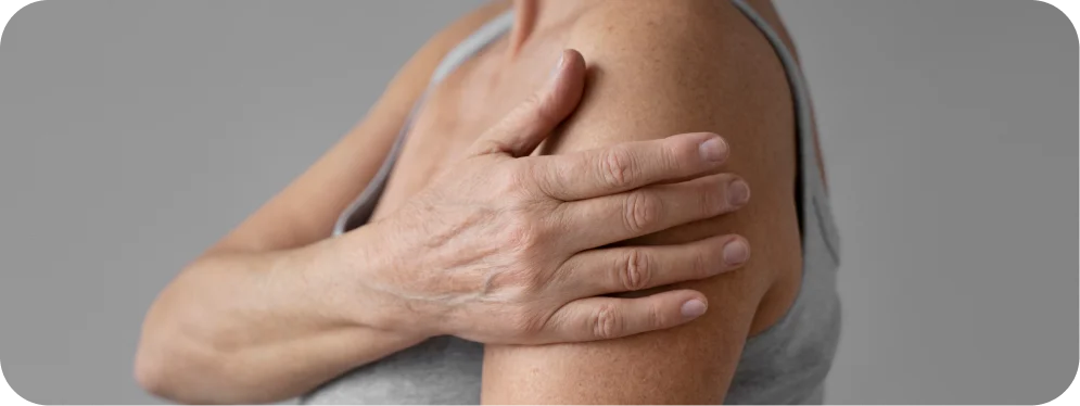
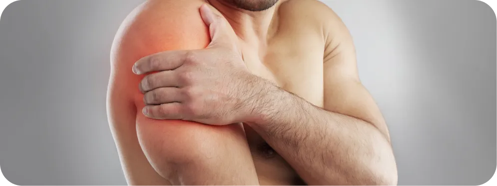
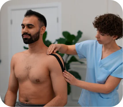
If you’ve recently been diagnosed with an AC joint injury, you may be feeling a mix of concern and uncertainty. These injuries—whether acute from a fall or sports incident, or chronic due to wear and tear—can significantly affect your shoulder function and quality of life. Understanding your diagnosis early and exploring your treatment options can make a huge difference in recovery and long-term shoulder health.
In this guide, we’ll walk you through the symptoms, diagnosis, and treatment options for AC joint injuries, with a special focus on Arthroscopic vs. Open AC Joint Repair/Reconstruction, helping you feel informed and confident about the next steps.
[tabla_contenidos]
What Are AC Joint Injuries (Acute or Chronic)?
The acromioclavicular (AC) joint is located at the top of your shoulder, where the collarbone (clavicle) meets the highest point of the shoulder blade (acromion). It plays an essential role in shoulder movement, stability, and overall upper-body function.
AC joint injuries occur when this joint is stressed, either suddenly (acute) or gradually (chronic):
Acute injuries usually result from a fall directly onto the shoulder, sports collisions, or accidents.
Chronic injuries develop over time due to repetitive stress, degenerative changes, or arthritis.
Recognizing the injury early is important because untreated AC joint damage can lead to persistent pain, shoulder weakness, and reduced range of motion.
Symptoms of AC Joint Injuries
The symptoms of an AC joint injury can vary depending on severity, but most patients notice some combination of the following:
Pain at the top of the shoulder, often worsening with overhead activities or lifting.
Visible bump or swelling over the AC joint in acute injuries.
Tenderness to touch along the joint line.
Reduced range of motion or difficulty performing daily tasks like reaching overhead or behind the back.
Shoulder weakness or instability, particularly in chronic injuries.
If these symptoms persist or worsen, it’s important to seek evaluation by an orthopedic specialist to prevent long-term complications.
How AC Joint Injuries Are Diagnosed
Diagnosing an AC joint injury typically involves a combination of physical examination and imaging:
Physical Exam:
Your doctor will assess tenderness, swelling, and any abnormal mobility in the AC joint.
Specific tests may be performed to evaluate ligament stability and pain with shoulder movement.
Imaging Studies
X-rays help identify fractures, joint separation, or degenerative changes.
MRI may be recommended in chronic or complex cases to evaluate soft tissues, ligaments, and cartilage.
A thorough diagnosis allows your orthopedic specialist to determine whether non-surgical care is appropriate or if surgical intervention is needed.
Treatment Options for AC Joint Injuries
Non-Surgical Treatments
Not all AC joint injuries require surgery. Mild to moderate injuries are often managed conservatively with:
Rest and activity modification to avoid movements that aggravate the joint.
Ice or heat therapy to reduce pain and inflammation.
Physical therapy focused on strengthening shoulder muscles and improving range of motion.
Anti-inflammatory medications for short-term pain relief.
While these approaches can be effective for many patients, severe acute injuries or chronic degenerative AC joint problems may benefit more from surgical repair or reconstruction.
Arthroscopic vs. Open AC Joint Repair/Reconstruction
When non-surgical treatments are insufficient, surgical intervention may be recommended to restore joint stability, reduce pain, and improve shoulder function. Two main approaches are used: arthroscopic and open AC joint repair/reconstruction.
How It Works
Arthroscopic AC Joint Repair:
Performed using small incisions and a tiny camera (arthroscope).
Allows the surgeon to precisely repair damaged ligaments, remove bone spurs, and reconstruct the joint with minimal disruption to surrounding tissues.
Often involves using specialized devices to stabilize the clavicle.
Open AC Joint Reconstruction:
Involves a slightly larger incision to directly access the AC joint.
Allows for reconstruction of torn ligaments and restoration of joint alignment, sometimes using grafts from your own tissue or synthetic materials.
Both procedures aim to restore shoulder stability, reduce pain, and prevent future joint degeneration.
When Surgery Is Recommended
Surgical repair is typically considered when:
The AC joint injury is severe or high-grade, with visible displacement of the collarbone.
Chronic pain or instability persists despite conservative therapy.
The patient is physically active or has high demands on the shoulder for work or sports.
Benefits and Success Rates
Arthroscopic repair offers smaller incisions, less soft tissue trauma, and faster early recovery.
Open reconstruction may provide robust repair in complex cases or when tissue quality is poor.
Success rates for both approaches are high, with most patients experiencing significant pain relief, improved strength, and restoration of shoulder function. Studies show that 85–95% of patients achieve excellent long-term outcomes when treated by an experienced orthopedic specialist.
Take Action for Your Shoulder Health
AC joint injuries can be painful and limit your daily activities, but with expert evaluation and modern surgical techniques, most patients regain strength, stability, and confidence in their shoulder.
If you are experiencing persistent pain, reduced motion, or instability, consult an experienced orthopedic specialist who can guide you through the best treatment options, including Arthroscopic vs. Open AC Joint Repair/Reconstruction. Early intervention and expert care give you the best chance for a full, lasting recovery and a return to the activities you love.
Don’t wait—schedule a consultation today to take the first step toward a healthy, pain-free shoulder.
A Pectoralis tendon rupture is a serious shoulder injury that can affect strength, function, and overall quality of life. While not as common as rotator cuff injuries, it is often seen in adults who engage in heavy lifting, weightlifting, or contact sports. Understanding the injury early is crucial, as timely treatment—especially surgical repair—can restore shoulder strength and function effectively.
If you’ve recently been diagnosed with a pectoralis tendon rupture, this guide will help you understand what it means, how it’s diagnosed, and why Open Pectoralis Tendon Repair is considered the gold standard for lasting recovery.
[tabla_contenidos]
Symptoms of a Pectoralis Tendon Rupture
A pectoralis tendon rupture typically presents with clear, noticeable symptoms. Many patients report:
Sudden pain in the chest or shoulder: Often described as a tearing or popping sensation during activity.
Bruising and swelling: Discoloration may develop along the chest or upper arm shortly after the injury.
Loss of strength: Difficulty pushing, lifting, or performing overhead activities.
Visible deformity: The chest or upper arm may appear sunken or asymmetric compared to the other side.
Tenderness: The area near the upper arm or chest can be very sensitive to touch.
Recognizing these signs early is important. Ignoring them may lead to chronic weakness, limited function, or cosmetic deformities.
How a Pectoralis Tendon Rupture is Diagnosed
Diagnosing a pectoralis tendon rupture involves a combination of physical examination and imaging studies.
Physical Examination
During your appointment, an orthopedic specialist will:
Assess range of motion and strength in your shoulder and chest.
Look for asymmetry in muscle contour.
Palpate for tenderness or gaps along the tendon.
Imaging Tests
To confirm the diagnosis and plan treatment, your doctor may recommend:
MRI (Magnetic Resonance Imaging): Provides detailed images of the tendon and surrounding tissues, showing the exact location and severity of the tear.
Ultrasound: Useful for assessing tendon integrity in real-time.
X-rays: While they don’t show tendons, they can rule out bone injuries that sometimes occur alongside tendon tears.
With a precise diagnosis, your orthopedic specialist can recommend the most effective treatment approach.
Treatment Options for Pectoralis Tendon Rupture
Treatment depends on the severity of the tear, your activity level, and your personal goals. Options typically include non-surgical care or surgical repair.
Non-Surgical Treatment
For partial tears or patients with lower physical demands, non-surgical management may be considered. This typically includes:
Rest and activity modification: Avoid lifting, pushing, or overhead activities.
Physical therapy: Focuses on strengthening surrounding muscles to compensate for some loss of function.
Pain management: Anti-inflammatory medications or short-term use of ice and heat therapy.
While non-surgical care can relieve pain and maintain some function, it often cannot restore full strength or correct cosmetic deformity. For most active adults, especially athletes or manual laborers, surgical repair is the recommended solution.
Open Pectoralis Tendon Repair: The Gold Standard
When it comes to restoring strength, function, and appearance, Open Pectoralis Tendon Repair is the most reliable treatment for a complete pectoralis tendon rupture.
What is Open Pectoralis Tendon Repair?
This is a surgical procedure in which your orthopedic surgeon:
Makes a small incision near the shoulder or chest.
Locates the torn tendon and carefully reattaches it to the bone using strong sutures or anchors.
Ensures the tendon is securely fixed in its natural position, allowing it to heal properly.
This approach is considered gold standard because it directly addresses the tear, restores normal anatomy, and provides predictable long-term results.
When is Surgery Recommended?
Open Pectoralis Tendon Repair is usually recommended for:
Complete tendon ruptures.
Active individuals or athletes who require full shoulder strength.
Patients seeking cosmetic correction of chest deformity.
Those who do not improve with non-surgical care.
Surgery is ideally performed within a few weeks of the injury for optimal outcomes.
Benefits of Open Pectoralis Tendon Repair
High success rate: Studies show most patients regain full or near-full strength after surgery.
Restored function: Patients can return to weightlifting, sports, and daily activities.
Cosmetic improvement: Corrects visible deformity of the chest and upper arm.
Long-term durability: Surgical repair significantly reduces the risk of chronic weakness or re-injury.
Choosing an Experienced Orthopedic Specialist
The success of Open Pectoralis Tendon Repair depends heavily on the surgeon’s experience and expertise. An orthopedic specialist with advanced training in shoulder and joint injuries can:
Precisely diagnose the injury.
Recommend the best treatment plan for your lifestyle.
Guide you through a safe, structured recovery.
Maximize the likelihood of a full return to activity.
Choosing a specialist you trust ensures that your repair is done with care, precision, and the best possible outcomes.
Take the Next Step Toward Recovery
If you suspect or have been diagnosed with a Pectoralis tendon rupture, it’s important to act promptly. Early evaluation by an orthopedic specialist can make a significant difference in your strength, function, and long-term satisfaction.
With Open Pectoralis Tendon Repair, most patients regain nearly full strength, restore chest symmetry, and confidently return to their favorite activities. Don’t wait—schedule a consultation today to discuss your options and take the first step toward a complete, lasting recovery.
A meniscus tear is one of the most common knee injuries affecting adults under 65, whether you’re an active athlete or someone who simply enjoys staying fit. Understanding your diagnosis early is crucial, not only to relieve pain but also to preserve long-term knee health.
In this article, we’ll walk you through the symptoms, how doctors diagnose meniscus tears, and the latest treatment options, including advanced surgical techniques designed to restore function and mobility.
[tabla_contenidos]
What is a Meniscus Tear?
The meniscus is a crescent-shaped cartilage in your knee that acts as a cushion between your thigh bone (femur) and shinbone (tibia). Each knee has two menisci—medial (inside) and lateral (outside)—which help absorb shock, stabilize the joint, and protect cartilage from wear.
A meniscus tear occurs when this cartilage is damaged or torn, often due to twisting, sudden movements, or gradual degeneration over time. Left untreated, a tear can lead to persistent pain, swelling, and eventually early osteoarthritis.
Symptoms of a Meniscus Tear
Patients with meniscus tears typically report the following signs:
Knee pain, often along the joint line.
Swelling or stiffness, which can worsen over a few hours or days.
Catching or locking, where the knee feels stuck or snaps during movement.
Difficulty bending or straightening the knee fully.
A feeling of instability, as if the knee might “give way.”
While some tears cause immediate pain after an injury, others develop gradually, particularly in older adults or those with degenerative changes.
How is a Meniscus Tear Diagnosed?
An accurate diagnosis is the first step toward effective treatment. Orthopedic specialists typically use a combination of:
Medical history and physical examination
Doctors assess pain location, range of motion, and perform specific maneuvers (like the McMurray or Apley tests) to detect meniscus damage.
Imaging studies
MRI scans are the gold standard for visualizing meniscus tears, showing the type, size, and location of the injury.
X-rays may be used to rule out bone injuries or arthritis but do not show cartilage damage.
Arthroscopy (Rare cases)
A minimally invasive procedure used for both diagnosis and treatment, particularly if imaging results are unclear.
Treatment Options for Meniscus Tears
Treatment depends on the type, size, and location of the tear, as well as the patient’s age, activity level, and overall knee health.
Non-Surgical Approaches
For partial tears or patients with lower physical demands, non-surgical management may be considered. This typically includes:
Rest and activity modification to reduce stress on the knee.
Ice and anti-inflammatory medications to control pain and swelling.
Physical therapy to strengthen surrounding muscles, improve flexibility, and stabilize the joint.
While non-surgical options can provide symptom relief, they do not repair the meniscus, leaving the joint at risk for further injury and long-term degeneration.
Surgical Options: How the Circumferential Stitch Restores Knee Function vs. Meniscectomy
When a meniscus tear is significant or symptoms persist, surgery may be recommended. The two main approaches are:
Meniscectomy
A meniscectomy involves removing the torn portion of the meniscus.
Pros:
Provides quick pain relief.
Effective for tears that cannot be repaired..
Cons:
Removes part of the meniscus, reducing cushioning in the knee.
Increases the risk of developing early arthritis.
Long-term knee function may be limited, especially for active adults.
Arthroscopic Meniscus Repair with Circumferential Stitch
The Circumferential Stitch, developed by Dr. Justin Saliman, focuses on repairing and preserving the meniscus rather than removing it.
How it works:
The technique encircles the tear with strong sutures, providing uniform compression along the edges.
This encourages healing even in areas with poor blood supply, which traditionally have a low chance of recovery.
Maintains the anatomical shape and stability of the meniscus, keeping the knee naturally cushioned and functional.
Benefits of the Circumferential Stitch:
Preserves more meniscus tissue, which protects the joint and reduces the risk of future degeneration.
Supports natural healing, allowing the meniscus to repair itself rather than simply removing damaged tissue.
Maintains knee stability and cushioning, which is essential for daily activities, sports, and long-term joint health.
Reduces the risk of early arthritis, making it a better option for younger, active adults who want to maintain knee function for years to come.
High success rates, even for complex tears that are difficult to repair using traditional techniques.
Why it’s generally better than meniscectomy:
While meniscectomy can provide faster short-term relief, it sacrifices meniscus tissue, which is crucial for long-term joint health. In contrast, Arthroscopic Meniscus Repair with the Circumferential Stitch not only addresses the immediate problem but also protects your knee for the future, preserving function, stability, and comfort.
In summary, whenever the meniscus can be repaired, this advanced repair technique is the preferred choice, offering patients the best combination of healing, joint preservation, and long-term knee health.
Why Choose Repair Over Removal?
Preserving your meniscus whenever possible is the best strategy for long-term knee health. While meniscectomy may offer faster initial relief, it sacrifices cartilage, which increases the likelihood of future arthritis. Arthroscopic repair with the Circumferential Stitch maximizes healing potential, stabilizes the knee, and protects your joint for years to come.
Take Action for Your Knee Health
If you have been diagnosed with a meniscus tear, consulting an experienced orthopedic specialist is crucial. They can evaluate your tear, explain the best surgical or non-surgical options, and help you choose a treatment plan that balances pain relief, joint preservation, and long-term function.
Modern techniques like the Circumferential Stitch in Arthroscopic Meniscus Repair offer patients the opportunity to heal naturally, maintain knee stability, and return confidently to the activities they love. Don’t wait—early intervention can make all the difference in protecting your knees for years to come.
By choosing a surgeon with expertise in this procedure, you can benefit from:
Preserving your meniscus to maintain cushioning and stability.
Promoting natural healing, even in challenging or complex tears.
Reducing the risk of future knee problems, such as early arthritis.
A personalized treatment plan tailored to your activity level, age, and lifestyle.
A meniscus root tear is a serious injury that affects the root attachment of the meniscus, the C-shaped cartilage in your knee that acts as a natural shock absorber. Unlike other meniscus tears, a root tear compromises the stability and function of the entire meniscus. This type of injury can dramatically alter the biomechanics of your knee, increasing stress on the joint and accelerating cartilage wear.
If left untreated, a meniscus root tear can lead to early-onset arthritis, joint degeneration, and long-term disability. That’s why it’s crucial to understand this condition early and explore the most effective treatment options.
[tabla_contenidos]
How a Meniscus Root Tear Feels
Recognizing the signs of a meniscus root tear can help you seek treatment before further damage occurs. Common symptoms include:
Pain around the inner or outer side of the knee: Pain often intensifies with twisting motions, squatting, or climbing stairs.
Swelling or stiffness: Swelling may appear gradually after activity, and stiffness can make it difficult to fully bend or straighten the knee.
Knee instability: You might feel the knee giving way or unable to fully support your weight, especially during sudden movements.
Catching, locking, or popping sensations: Particularly with complex tears, the knee may momentarily catch or lock, causing discomfort or limiting movement.
Reduced activity tolerance: Patients often notice they cannot perform routine activities like walking long distances, kneeling, or sports without pain.
These symptoms vary based on activity level, age, and whether the tear was caused by trauma or degenerative changes over time. Understanding your symptoms is the first step toward effective treatment.
How Doctors Confirm a Meniscus Root Tear
Accurate diagnosis of a meniscus root tear requires a combination of physical examination, imaging, and clinical assessment.
Physical Examination: Your orthopedic specialist will assess the knee’s range of motion, stability, and tenderness points. Specific maneuvers can indicate a root tear, such as tests for joint line pain and rotational instability.
MRI (Magnetic Resonance Imaging): MRI is the most reliable tool for visualizing soft tissue structures like the meniscus. It can confirm the location, size, and severity of the tear.
X-Rays: While X-rays do not show the meniscus itself, they help rule out other causes of knee pain and can detect early signs of joint degeneration or arthritis.
Patient History: Your doctor will evaluate how the injury occurred, whether suddenly or gradually, and discuss your activity level, previous knee injuries, and current symptoms.
Prompt and accurate diagnosis is critical because untreated root tears can lead to worsening joint instability, faster cartilage wear, and increased risk of developing osteoarthritis.
Treatment Options
Non-Surgical Approaches
Some patients with minimal symptoms or lower activity demands may benefit from non-surgical treatment. These approaches focus on managing pain, reducing inflammation, and maintaining mobility, but they do not repair the root of the meniscus. Common non-surgical options include:
Activity modification: Avoiding deep squatting, twisting, or high-impact movements.
Physical therapy: Strengthening the quadriceps, hamstrings, and supporting muscles to stabilize the knee.
Pain and inflammation management: Over-the-counter medications or targeted injections may help relieve symptoms temporarily.
It’s important to note that while these measures can reduce discomfort, they do not restore knee stability or prevent long-term joint degeneration.
Arthroscopic Meniscus Root Repair with Circumferential Stitch
For most patients with a meniscus root tear, surgical repair is the most effective solution. Dr. Justin Saliman has pioneered the Arthroscopic Meniscus Root Repair technique using the Circumferential Stitch, often facilitated with the NovoStitch device. This approach focuses on preserving the meniscus, restoring natural knee mechanics, and protecting joint health.
How It Works
Minimally invasive arthroscopy: Small incisions allow the surgeon to access the meniscus without disrupting surrounding tissue, reducing overall trauma to the knee.
Circumferential Stitch technique: This technique encircles the meniscus root with strong sutures, creating uniform compression across the tear to optimize healing.
NovoStitch device: This specialized tool allows precise placement of sutures even in hard-to-reach areas, ensuring a secure, anatomically correct repair.
Meniscus preservation: Unlike traditional meniscectomy, which removes damaged tissue, this technique focuses on healing and stabilizing the meniscus to maintain its natural cushioning function.
Benefits for Patients
Patients who undergo Arthroscopic Meniscus Root Repair with Circumferential Stitch experience multiple advantages:
Preserves natural knee function: Repairing the root maintains the meniscus’ role as a shock absorber, protecting cartilage from excessive wear.
Restores stability: By reattaching the root, the knee regains proper biomechanics, which prevents giving way and improves confidence in daily activities.
High success rates: Clinical studies and surgical experience demonstrate consistent healing, even for complex or degenerative tears.
Durable results: Unlike partial meniscectomy, which may provide temporary relief, repair addresses the root cause of instability, offering long-term joint protection.
Enhanced activity potential: Patients can often return to low- and moderate-impact activities with restored knee function and comfort.
Protection against future surgeries: Maintaining the meniscus reduces the likelihood of partial or total knee replacement later in life.
Tailored for active lifestyles: Athletes or active adults benefit from a repair that supports performance while minimizing the risk of re-injury.
This procedure provides a comprehensive solution that focuses on the patient’s long-term joint health, emphasizing healing, stability, and functional improvement over simple tissue removal.
Comparing Repair vs. Meniscectomy
Unlike meniscectomy, which removes damaged tissue, the Circumferential Stitch repair:
Preserves meniscus volume and joint cushioning.
Maintains stability and prevents abnormal load distribution.
Reduces the risk of accelerated osteoarthritis.
Provides superior functional outcomes for patients of all activity levels.
In most cases, repair is strongly preferred over removal, especially in patients who wish to maintain active lifestyles and protect their knees long-term.
Take Action for Your Knee Health
If you’ve been diagnosed with a meniscus root tear, early intervention is key to protect your knee and maintain long-term mobility. Seeking care from an experienced orthopedic specialist ensures that your knee is evaluated thoroughly and that you receive the most effective treatment tailored to your needs.
By consulting with a specialist, you can benefit from:
Preservation of the meniscus and protection of your joint.
Restoration of knee stability and natural mechanics.
Reduced risk of early arthritis and future joint problems.
Personalized treatment plans designed to fit your activity level and lifestyle.
Don’t wait until pain or instability limits your daily activities. Schedule a consultation with an orthopedic specialist to discuss your options and take the first step toward a healthier, stronger knee.
Your meniscus plays a crucial role in keeping your knee healthy and pain-free. These two C-shaped pieces of cartilage act as shock absorbers between your thigh bone and shin bone, helping distribute weight evenly during walking, running, or squatting.
Meniscus extrusion happens when part of the meniscus is pushed—or “extruded”—out of its normal position along the edge of the tibia (shinbone). This may sound like a small detail, but it can have a big impact. When the meniscus moves out of place, it can no longer protect the joint properly, leading to pain, swelling, cartilage damage, and eventually early arthritis if left untreated.
Understanding this condition early and seeking the right treatment is key to protecting your knee and preventing long-term damage.
[tabla_contenidos]
Common Symptoms of Meniscus Extrusion
Many patients with meniscus extrusion notice gradual discomfort rather than a sudden injury. The condition often develops alongside degenerative meniscus tears or after previous knee trauma.
Typical Symptoms Include:
Pain along the joint line, especially on the inner (medial) side of the knee.
Swelling or stiffness after activity.
Clicking or catching sensations inside the knee.
Feeling of instability or “giving way.”
Limited range of motion, particularly difficulty squatting or twisting.
In some cases, the pain may seem mild at first but worsens over time as the meniscus continues to lose its normal alignment and cushioning ability.
How Meniscus Extrusion Is Diagnosed
Diagnosing meniscus extrusion usually starts with a careful clinical examination by an orthopedic specialist, followed by imaging to confirm the extent of displacement.
Physical Examination
Your doctor will assess for tenderness along the joint line, swelling, and mechanical symptoms. They’ll also check your knee’s stability and alignment.
MRI Scan
MRI is the gold standard for confirming meniscus extrusion. It shows how far the meniscus has shifted beyond the edge of the tibial plateau.
A displacement of more than 3 mm is typically considered significant.
MRI can also reveal any associated meniscal tears or cartilage damage.
Understanding these findings helps determine whether conservative care or surgery offers the best chance for long-term knee preservation.
Treatment Options for Meniscus Extrusion
Treatment depends on the severity of the extrusion, associated meniscus tears, and how much pain or functional limitation the patient is experiencing.
Non-Surgical Management (Early or Mild Cases)
For minor extrusion without mechanical symptoms or cartilage injury, doctors may recommend:
Activity modification: avoiding deep squats, twisting, or high-impact exercises.
Physical therapy: strengthening surrounding muscles to support knee stability.
Anti-inflammatory medications or corticosteroid injections to manage pain and swelling.
Bracing: in selected cases, to offload stress from the affected compartment.
While these approaches can help reduce discomfort, they don’t correct the underlying structural problem—the displaced meniscus still remains out of position and unable to protect the cartilage effectively.
That’s why, for persistent pain or progressive extrusion, surgical repair may be the best solution.
When the meniscus is extruded and no longer anchored properly to the tibia, surgeons can now restore its natural position and function through a procedure known as Arthroscopic Meniscus Centralization (also called Meniscotibial Ligament Repair).
This innovative, minimally invasive technique has transformed how orthopedic specialists manage meniscus extrusion.
What Is Meniscus Centralization?
In a healthy knee, the meniscotibial ligament secures the meniscus tightly to the edge of the tibia. When this ligament becomes loose or torn, the meniscus slips outward—causing extrusion.
Meniscus centralization uses arthroscopic (camera-guided) tools to re-anchor the meniscus back to the tibia, restoring its natural contour and ability to distribute load evenly across the joint.
How the Procedure Works
During surgery:
Small arthroscopic incisions are made around the knee.
Specialized suture anchors are placed along the tibial edge.
The surgeon repositions the meniscus and secures it using circumferential stitches or all-suture anchors, reinforcing the meniscotibial ligament and pulling the meniscus back into its native position.
The stability and mobility of the meniscus are verified in real time before completing the procedure.
This approach is often combined with meniscus repair if there is a concurrent tear, ensuring full restoration of the meniscus structure.
Benefits of Arthroscopic Meniscus Centralization
Meniscus centralization addresses the root cause of pain and joint degeneration by restoring the normal biomechanics of the knee rather than merely managing symptoms.
Preserves the native meniscus, maintaining knee shock absorption and stability.
Minimally invasive — small incisions, less pain, faster rehabilitation.
Improved long-term outcomes compared to partial meniscectomy (removal).
Reduced risk of future joint degeneration and need for knee replacement.
Studies and clinical experience have shown promising success rates, with high rates of meniscal healing and significant improvements in pain and function within months.
What Patients Can Expect After Surgery
After Arthroscopic Meniscus Centralization, patients usually follow a structured rehabilitation program tailored to protect the repair while restoring motion and strength.
Post-Operative Highlights:
Weight-bearing is gradually reintroduced, often with crutches and a brace during the first few weeks.
Physical therapy begins early, focusing on controlled range of motion and muscle activation.
As healing progresses, strength and balance training help patients return to walking, low-impact sports, and eventually full activity.
Every recovery journey is unique, but most patients report steady improvement in knee comfort and confidence as the meniscus heals and regains its stabilizing function.
The ultimate goal is joint preservation — protecting the meniscus so it can continue doing its vital job for years to come.
The Importance of Expert Care
Not every meniscus extrusion needs surgery, but when symptoms persist or imaging shows significant displacement, consulting an experienced orthopedic specialist is crucial.
Surgeons skilled in Arthroscopic Meniscus Centralization understand the delicate balance between joint mechanics and tissue preservation. Choosing this advanced approach can mean the difference between short-term relief and long-term joint health.
Take the Next Step Toward Knee Health
If you’ve been diagnosed with meniscus extrusion, don’t ignore your symptoms or settle for temporary fixes. Modern surgical techniques like Arthroscopic Meniscus Centralization (Meniscotibial Ligament Repair) offer real, lasting solutions — helping restore your knee’s natural alignment, stability, and comfort.
Talk to an orthopedic specialist who focuses on meniscus preservation and repair to explore the best treatment options for you. Taking action today can help you stay active, protect your cartilage, and keep your knees healthy for the long run.
Chondral defects, or injuries to the cartilage that covers the joints, are common yet often overlooked conditions that can lead to serious long-term consequences if not addressed early. Cartilage plays a critical role in allowing smooth and painless joint movement.
When this cartilage becomes damaged—whether due to trauma, wear and tear, or degeneration—it can significantly affect your mobility and quality of life. In this article, we’ll discuss what a chondral defect/injury is, how it’s diagnosed, and the most effective treatments available, including surgical options like Arthroscopic, Open Cartilage Restoration (Cartiheal), and Implantation (MACI).
[tabla_contenidos]
What Is a Chondral Defect/Injury?
A chondral defect is a type of cartilage injury that affects the smooth, elastic tissue lining your joints, especially the knee, hip, and ankle. This cartilage, known as articular cartilage, is vital for reducing friction between bones and absorbing shock during movement. When it becomes damaged, the bones in the joint rub against each other, causing pain, stiffness, and potentially leading to further joint deterioration.
Chondral defects can be caused by a variety of factors, including:
Trauma or injury: A direct blow or sports-related injury, like twisting the knee, can cause cartilage damage.
Wear and tear (Osteoarthritis): Age and repetitive movements can gradually wear down cartilage, making it more susceptible to injury.
Overuse: Frequent high-impact activities or repetitive stress can lead to cartilage degradation over time.
If left untreated, a chondral defect can evolve into more severe conditions, such as osteoarthritis, leading to chronic pain, limited range of motion, and even permanent joint damage.
Symptoms of Chondral Defect/Injury
The symptoms of a chondral defect can vary depending on the location and severity of the injury. However, the most common signs include:
Pain: You might feel pain around the affected joint, especially when performing certain movements or after physical activity.
Swelling: The injured area may swell up, which is a result of inflammation in response to the damage.
Stiffness: You may experience a feeling of tightness or decreased range of motion in the joint, particularly after periods of rest.
Locking or catching: In some cases, damaged cartilage can cause the joint to “catch” or “lock,” making it difficult to move the joint properly.
Instability: The joint might feel unstable or weak, as if it could “give way” under pressure.
If you notice these symptoms, it’s important to seek medical attention early to prevent the condition from worsening and to explore treatment options that can relieve pain and restore function.
How Is Chondral Defect/Injury Diagnosed?
Diagnosing a chondral defect involves a thorough examination by an orthopedic specialist. Here’s what to expect during the diagnostic process:
Physical Exam
Your doctor will first perform a physical exam to assess the affected joint’s range of motion, tenderness, and any signs of instability. They may ask you about any recent injuries, physical activities, or family history of joint issues, such as osteoarthritis.
Imaging Tests
To confirm the diagnosis, your doctor will likely order imaging tests, including:
X-rays: While X-rays can’t directly show cartilage damage, they help rule out other causes of pain, such as fractures or joint misalignment.
MRI (Magnetic Resonance Imaging): This is the most accurate imaging technique for assessing cartilage damage. An MRI creates detailed images of both soft tissues and bone, allowing your doctor to see the extent of the chondral defect.
Arthroscopy: In some cases, a minimally invasive procedure called arthroscopy might be performed to visualize the damage directly inside the joint. This involves inserting a small camera (arthroscope) through a tiny incision.
Once the extent of the cartilage damage is confirmed, your doctor will discuss treatment options based on the severity of the injury and your personal activity level.
Treatment Options for Chondral Defect/Injury
The treatment for a chondral defect will depend on several factors, including the size and location of the injury, your age, activity level, and overall health. There are both non-surgical and surgical treatment options available.
Non-Surgical Treatments
For minor chondral defects, non-surgical treatments may be recommended to reduce symptoms and promote healing. These can include:
Rest and Activity Modification: Limiting high-impact activities to avoid further stress on the joint.
Physical Therapy: Targeted exercises to strengthen the muscles around the joint, improve stability, and reduce strain on the cartilage.
Medications: Nonsteroidal anti-inflammatory drugs (NSAIDs) can help manage pain and inflammation.
Injections: Corticosteroid injections or hyaluronic acid injections may be used to reduce inflammation and improve joint lubrication.
If non-surgical treatments don’t provide sufficient relief, surgical options might be considered. Below, we will focus on three primary surgical procedures: Arthroscopic Surgery, Open Cartilage Restoration (Cartiheal), and Implantation (MACI).
Surgical Treatment Options
When non-surgical treatments aren’t enough to resolve a chondral defect, surgical intervention becomes necessary. Depending on the severity of the cartilage damage, different procedures may be recommended. Below, we will discuss three common surgical treatments for chondral defects: Arthroscopic Surgery, Open Cartilage Restoration (Cartiheal), and Implantation (MACI).
Arthroscopic Surgery
How It Works
Arthroscopic surgery is a minimally invasive procedure that involves small incisions and the use of a tiny camera (arthroscope) to view and treat the cartilage damage. This approach is ideal for smaller defects or when the damage is located in areas that are easy to access.
Cartilage Debridement and Microfracture: During the procedure, the surgeon may perform “debridement,” which involves cleaning the damaged area by removing any loose cartilage or debris. In cases where the damage is deeper, the surgeon may use a technique called microfracture, where tiny holes are created in the bone to stimulate the growth of new cartilage.
Shaping and Smoothing: Sometimes, the surgeon will also smooth or reshape the cartilage to ensure that the joint moves as fluidly as possible. This can improve function and reduce pain.
When It’s Recommended
Arthroscopic surgery is typically recommended for patients with smaller cartilage defects, especially when the damage is confined to one specific area. It is also the go-to treatment for individuals with minimal joint degeneration who want to preserve as much natural cartilage as possible.
Success Rates
Arthroscopic surgery has a high success rate for small defects, with studies showing that most patients experience significant pain relief and functional improvement. The minimally invasive nature of the procedure, combined with short recovery times, makes it a popular option for many patients. However, larger defects or deep cartilage injuries may not be fully addressed with arthroscopy alone, which is why additional treatments might be considered.
Open Cartilage Restoration (Cartiheal)
How It Works
Cartiheal is a relatively new and innovative surgical approach designed to treat larger or deeper cartilage defects. It involves the implantation of a bioactive collagen scaffold, which serves as a foundation for new cartilage growth. This technique encourages the body’s natural healing process and allows the joint to regenerate its own cartilage over time.
Bioactive Collagen Scaffold: The procedure involves placing a collagen scaffold into the damaged area of the cartilage. This scaffold is impregnated with growth factors that stimulate the body’s ability to heal itself by promoting the regeneration of cartilage cells.
Bone and Cartilage Integration: The scaffold integrates with the surrounding bone and cartilage to form a stable foundation, encouraging natural healing and minimizing the risk of future damage. Over time, the body gradually replaces the scaffold with its own new cartilage.
When It’s Recommended
Cartiheal is often recommended for patients with larger chondral defects or those whose injuries are too extensive for traditional arthroscopic surgery. It’s also a great option for individuals who have previously undergone arthroscopic surgery but are still dealing with persistent cartilage damage.
Success Rates
The Cartiheal procedure has shown promising results, particularly for patients with medium-to-large defects. Studies have reported excellent cartilage regeneration and improvements in joint function and pain reduction. The success of this procedure is largely due to its ability to regenerate cartilage using the body’s own cells and natural healing mechanisms.
Implantation (MACI)
How It Works
Matrix-induced Autologous Chondrocyte Implantation (MACI) is an advanced technique for repairing larger cartilage defects, and it involves using your own cartilage cells to create new cartilage. Here’s how it works:
Harvesting Cartilage Cells: The first step in the MACI procedure involves collecting healthy cartilage cells from an area of your joint that is less weight-bearing. These cells are sent to a lab, where they are cultured and multiplied to create a larger supply.
Cell Implantation: Once the cells are ready, they are implanted onto a biodegradable scaffold and inserted into the damaged cartilage area. The scaffold provides support for the new cells and helps them integrate into the joint over time.
When It’s Recommended
MACI is most beneficial for individuals with significant cartilage damage in weight-bearing joints, such as the knee, where smaller surgical options may not be effective. It is typically recommended for patients who have not responded to other treatments, including arthroscopic surgery or microfracture.
Success Rates
MACI offers excellent long-term outcomes, particularly for those with large defects or significant cartilage loss. Studies have shown high success rates, with most patients experiencing substantial improvements in joint function, pain relief, and a significant reduction in the need for further surgeries. The procedure’s success is largely due to its ability to create biologically integrated, durable cartilage that mimics natural tissue.
What to Expect After Surgery
The recovery process for cartilage restoration surgery varies depending on the procedure, the extent of cartilage damage, and the patient’s individual health. Here’s what you can generally expect after each type of surgery:
Arthroscopic Surgery
With arthroscopic surgery, many patients experience significant relief from pain, improved joint function, and enhanced mobility. However, the results can vary depending on the size and location of the defect.
Open Cartilage Restoration (Cartiheal)
Cartiheal can be highly effective for larger defects, with many patients reporting excellent pain reduction and functional recovery in the long term.
Implantation (MACI)
MACI is a great option for large cartilage defects, and patients often report substantial improvements in joint function and pain relief. As the body gradually integrates the new cartilage, long-term benefits continue to improve.
By taking a comprehensive approach to both the surgery and recovery phases, patients can achieve the best possible outcome. Each treatment offers distinct advantages based on the severity of the injury, and understanding what to expect at each stage of recovery is crucial for long-term success.
Why Choose Expert Care
Chondral defects can be challenging, but with the right treatment, many patients experience a significant improvement in their joint function and quality of life. Whether you’re exploring non-surgical treatments or considering surgical options like Arthroscopic Surgery, Cartiheal, or MACI, it’s essential to consult with an experienced orthopedic specialist who can guide you through the most appropriate course of action.
Taking the first step toward treatment now can prevent further joint damage and improve your overall health. If you’ve been diagnosed with a chondral defect, don’t wait—schedule a consultation today to learn how we can help restore your joint function and relieve your pain.
When knee pain or swelling doesn’t go away, especially after an injury, it can be frustrating — and even concerning. One possible cause is something called an Osteochondral lesion (also known as an Osteochondritis Dissecans, or OCD lesion).
Although the name sounds complicated, understanding this condition and its treatment options can help you feel more confident and in control of your recovery.
[tabla_contenidos]
What Is an Osteochondral/OCD Lesion?
An osteochondral lesion occurs when a small area of bone and its overlying cartilage inside a joint — most often the knee, but sometimes the ankle or elbow — becomes damaged.
This damage may happen after an injury, repetitive stress, or from reduced blood flow to the bone beneath the cartilage. Over time, this weak spot can cause pain, swelling, and even a small fragment of cartilage and bone to loosen inside the joint.
In younger, active adults under 65, it’s one of the most common causes of persistent joint pain, particularly after sports injuries or repeated impact activities.
Early diagnosis and treatment are crucial because cartilage doesn’t heal on its own — and when left untreated, the condition can worsen, increasing the risk of early arthritis.
Common Symptoms
Patients with osteochondral or OCD lesions usually report symptoms that can vary depending on the size and location of the lesion, but the most frequent include:
Deep, aching joint pain, often worsened by activity
Swelling or stiffness, especially after exercise
Locking or catching sensations in the joint
Feeling of instability or “giving way” during movement
Decreased range of motion or discomfort with weight-bearing
Many patients describe the pain as intermittent — good days and bad days — which often leads them to delay seeking care. However, early evaluation by an orthopedic specialist can make a big difference in outcomes.
How Is an Osteochondral/OCD Lesion Diagnosed?
Diagnosis typically starts with a thorough physical examination. Your orthopedic surgeon will assess swelling, tenderness, range of motion, and joint stability.
Imaging Studies
To confirm the diagnosis and determine the extent of damage, several imaging tools are used:
X-rays – to visualize bone alignment and rule out fractures
MRI scans – to see the cartilage and bone in detail, and to evaluate if the fragment is still attached or loose
CT scans – sometimes used to assess bone healing or plan surgery
These tests help the surgeon determine whether the lesion is stable (attached) or unstable (loose or displaced) — a key factor in deciding whether surgery is necessary.
Treatment Options for Osteochondral/OCD Lesions
Treatment depends on several factors — including lesion size, stability, age, and activity level.
Non-Surgical Management
For small, stable lesions, especially in younger patients, non-surgical care may be tried first. This may include:
However, if symptoms persist or imaging shows an unstable fragment or larger cartilage defect, surgical intervention is often the best solution to restore joint health and prevent further degeneration.
Surgical Treatment Options
Arthroscopic vs. Open Osteochondral Pinning vs. Cartiheal or MACI
When surgery is needed, the goal is to restore the damaged cartilage and underlying bone, allowing for smooth joint motion and long-term function. Depending on the case, your surgeon may recommend one of the following procedures.
Arthroscopic Osteochondral Pinning
What it is
Arthroscopic pinning is a minimally invasive procedure performed through small incisions using a camera (arthroscope). The surgeon identifies the loose or unstable cartilage fragment and reattaches it to the bone using small, bioabsorbable pins or screws.
When it’s recommended
This approach is typically used when the osteochondral fragment is still viable (alive) and large enough to be fixed back into place, especially in younger or active adults.
Benefits
Minimally invasive — less pain and quicker healing
Preserves the patient’s natural cartilage
Small incisions and minimal scarring
High rates of successful healing when done early
Success rates
Studies show 80–90% success in fragment healing and pain relief, especially when performed before the cartilage deteriorates significantly.
Open Osteochondral Pinning (or Fixation)
What it is
In some cases, particularly when the lesion is large, deep, or difficult to access arthroscopically, an open approach is preferred. The surgeon makes a slightly larger incision to directly visualize the lesion, realign the bone and cartilage, and secure it using specialized bioabsorbable pins or small screws.
When it’s recommended
Large or complex lesions
Detached fragments that need precise repositioning
Failed prior arthroscopic fixation
Benefits
Direct visualization allows more accurate placement of the fragment
Reliable fixation for larger or irregular defects
Restores the native cartilage surface and joint function
Success rates
Most studies report 85–95% good to excellent outcomes, particularly when combined with proper rehabilitation and activity modification during recovery.
Cartiheal (Agili-C) or MACI (Matrix-Induced Autologous Chondrocyte Implantation)
For larger or more chronic defects where the cartilage is no longer viable, regenerative cartilage restoration procedures are an advanced solution.
Cartiheal (Agili-C):
Cartiheal uses a biological scaffold made of natural minerals that supports new bone and cartilage growth. It doesn’t require cell harvesting and can be used in both osteochondral and purely cartilage lesions.
MACI is a two-step regenerative procedure. In the first step, a small sample of your healthy cartilage cells is taken and grown in a lab. In the second step, these cells are implanted on a collagen membrane and placed over the damaged area to regrow cartilage naturally.
Benefits
Uses your own cells for long-term cartilage restoration
Restores smooth, functional cartilage surface
Especially useful for large or chronic defects
Success rates
Clinical trials report 80–90% good to excellent results, with long-term durability for 10+ years when proper rehabilitation is followed.
Long-Term Outlook
With modern surgical techniques such as Arthroscopic and Open Osteochondral Pinning, Cartiheal, and MACI, most patients experience significant improvement in joint function, reduced pain, and a return to normal activity — even competitive sports. The key to success is early diagnosis, appropriate surgical planning, and a guided rehabilitation program.
When to See an Orthopedic Specialist
If you’ve been diagnosed with an Osteochondral/OCD lesion or continue to experience knee pain that doesn’t improve with rest, consulting an experienced orthopedic surgeon is the best next step.
Prompt evaluation can prevent worsening damage and help you explore the most effective treatment — from minimally invasive pinning to advanced cartilage restoration procedures.
Take the First Step Toward Full Recovery
Living with an Osteochondral lesion doesn’t mean giving up the activities you love. With modern techniques like Arthroscopic vs. Open Osteochondral Pinning vs. Cartiheal or MACI, patients today have multiple effective options to restore their joint health and return to an active lifestyle — safely and confidently.
If you’re struggling with persistent knee pain or have recently been diagnosed, schedule a consultation with an orthopedic specialist to discuss your personalized treatment plan and take the first step toward lasting recovery.
A patellar tendon or quadriceps tendon rupture is a serious injury that affects the connection between your thigh muscles and your kneecap (patella), or between the kneecap and your shinbone (tibia). These tendons are essential for straightening the knee, walking, climbing stairs, and even standing up from a chair.
When one of these tendons tears, the leg can lose its ability to extend properly — making simple movements extremely difficult or even impossible. While this injury can happen to anyone, it most often affects active adults under 65 who experience a sudden jump, fall, or trauma to the knee. Early diagnosis and treatment are crucial to restore full knee function and avoid long-term complications like weakness, stiffness, or instability.
[tabla_contenidos]
How to Recognize a Patellar or Quadriceps Tendon Rupture
A tendon rupture in the front of the knee often presents very suddenly. Patients typically describe a sharp “pop” or tearing sensation, followed by immediate pain and difficulty moving the leg.
Common symptoms include:
Sudden, severe pain just above (quadriceps tendon) or below (patellar tendon) the kneecap
Swelling and bruising around the front of the knee
A visible gap or indentation where the tendon has torn
Inability to straighten the knee or lift the leg when lying down
Difficulty walking or bearing weight on the injured leg
The kneecap may appear higher or lower than normal, depending on which tendon is affected
If you experience these symptoms after a fall or sports injury, it’s important to seek medical attention right away. Early evaluation greatly improves the success of treatment and recovery.
How Doctors Confirm the Injury
Your orthopedic specialist will begin by asking about how the injury occurred and performing a physical examination. During the exam, your doctor will assess your ability to straighten the knee, check for swelling, and feel for any gaps in the tendon.
To confirm the diagnosis and determine the severity of the tear, imaging tests are usually ordered:
X-rays can show changes in the position of the kneecap (too high or too low).
MRI (Magnetic Resonance Imaging) is the most accurate test for identifying a partial or complete tendon rupture and assessing damage to surrounding structures.
Ultrasound may also be used to visualize the tendon tear in real-time.
Accurate diagnosis helps your surgeon decide whether non-surgical treatment is possible or if surgery is required to restore knee function.
Treatment Options for Patellar/Quad Tendon Rupture
Non-Surgical Treatment (For Partial Tears Only)
Partial tears or minor strains may sometimes be treated without surgery, especially if the tendon is still partially intact. Treatment usually includes:
Immobilization with a knee brace or cast to keep the leg straight
Physical therapy after several weeks to regain motion and strength
Gradual return to normal activities over 2–3 months
However, in most cases — particularly complete ruptures — non-surgical options cannot restore normal function. That’s when surgical repair becomes necessary.
Surgical Treatment: Open Patellar Tendon/Quad Tendon Repair
When the tendon is fully torn, the most effective solution is a surgical procedure called Open Patellar Tendon/Quad Tendon Repair. This operation directly repairs the torn tendon and restores the natural connection between your muscles, kneecap, and leg bone.
When Surgery Is Recommended
Surgery is strongly recommended when:
The tendon is completely ruptured (confirmed by MRI)
You are unable to straighten your leg
The kneecap is displaced from its normal position
You want to regain full knee function and return to sports or active work
Delaying surgery can make the repair more difficult, as the tendon and muscles may shorten or scar down. Ideally, the repair should be performed within a few weeks of injury for the best results.
How Open Patellar Tendon/Quad Tendon Repair Works
During this procedure, your orthopedic surgeon makes a small incision over the front of the knee to carefully expose the torn tendon. The damaged ends are identified and reattached using strong surgical sutures or anchors that secure the tendon back to the kneecap or bone.
Depending on the injury:
Patellar tendon repairs involve reattaching the tendon to the bottom of the kneecap.
Quadriceps tendon repairs involve reattaching the tendon to the top of the kneecap.
In some cases, if the tissue is weak or retracted, the surgeon may use reinforcement with grafts or synthetic materials to strengthen the repair. The procedure typically takes less than 90 minutes and is done under regional or general anesthesia.
Benefits of Open Patellar Tendon/Quad Tendon Repair
Patients can expect excellent outcomes with timely surgery and proper rehabilitation. The main benefits include:
Restoration of knee strength and stability
Improved mobility and ability to walk normally again
Low risk of re-rupture when performed by an experienced orthopedic surgeon
Return to sports and active lifestyle within several months
High patient satisfaction — most people regain nearly full range of motion
Clinical studies show that over 90% of patients recover good to excellent function after surgical repair when combined with structured rehabilitation.
Success Rates and Long-Term Outlook
The long-term outlook after Open Patellar Tendon/Quad Tendon Repair is excellent. When performed promptly and followed by a dedicated rehab program, the success rate exceeds 90%, and most patients return to their prior level of activity.
Complications such as stiffness, weakness, or re-rupture are uncommon and can usually be minimized through early motion, consistent physical therapy, and follow-up with your orthopedic specialist.
Take the First Step Toward Full Recovery
A patellar or quadriceps tendon rupture is a serious injury, but with expert surgical care, full recovery is absolutely achievable. The key is early diagnosis and timely treatment by an experienced orthopedic specialist.
If you’re struggling with knee pain, weakness, or difficulty straightening your leg after an injury, don’t wait — proper evaluation and treatment can make all the difference.
Schedule a consultation with an orthopedic surgeon to discuss whether Open Patellar Tendon/Quad Tendon Repair is right for you. With the right surgical approach and personalized rehabilitation, you can confidently return to an active, pain-free life.
If you’ve been diagnosed with medial compartment chondromalacia, you’re not alone — and the good news is, there are effective ways to manage and even restore knee function. Many adults under 65 experience this condition, especially those who are active or have had previous knee injuries.
The first step toward meaningful recovery is understanding the “why” behind your knee pain. In this article, we’ll explain what medial compartment chondromalacia is, what symptoms to watch for, how it’s diagnosed, and the most effective treatments available today — including a promising minimally invasive solution called the MISHA Knee Implantation.
[tabla_contenidos]
What Is Medial Compartment Chondromalacia?
“Chondromalacia” refers to the softening or deterioration of the cartilage that cushions the joint. In the medial compartment (the inner part of the knee), this means the smooth cartilage covering the ends of the femur and tibia starts to wear down.
Over time, this breakdown can cause friction, inflammation, and discomfort when walking, climbing stairs, or even standing for long periods.
Why Early Diagnosis Matters
Catching medial compartment chondromalacia early allows your orthopedic specialist to slow down or even reverse progression before the cartilage loss leads to arthritis or joint deformity. The earlier we act, the better the long-term outlook for joint preservation.
Common Symptoms
Patients often describe their knee pain as:
A dull, aching pain on the inner side of the knee
Stiffness or swelling, especially after activity or long periods of sitting
Grinding or clicking sensations when bending the knee
Pain when going up or down stairs
Loss of confidence in the knee — feeling like it may “give out”
In the early stages, symptoms may come and go. As the cartilage damage progresses, pain becomes more persistent and can start to interfere with everyday activities.
How Is Medial Compartment Chondromalacia Diagnosed?
An orthopedic evaluation begins with a detailed physical exam, during which your doctor checks for tenderness, range of motion, and joint stability.
Imaging Tests
To confirm the diagnosis and assess cartilage damage, imaging is typically performed:
X-rays: Show bone alignment and joint space narrowing.
MRI: Provides a clear view of cartilage, meniscus, and soft tissue health.
Diagnostic arthroscopy (in rare cases): A minimally invasive camera procedure to directly visualize the joint if needed.
These tests help your surgeon determine both the severity of cartilage wear and whether it’s isolated to the medial compartment or involves other areas of the knee.
Treatment Options for Medial Compartment Chondromalacia
Treatment depends on how advanced the cartilage wear is and how much it’s affecting your daily life.
Non-Surgical Treatments
In the early stages, many patients benefit from:
Activity modification: Avoiding deep squats, high-impact exercise, and repetitive kneeling.
Physical therapy: Focused on strengthening the quadriceps and improving knee alignment.
Anti-inflammatory medications or injections: Reduce pain and swelling.
Weight management: Reduces stress on the inner knee joint.
However, when pain persists despite these measures — and imaging shows cartilage loss in the medial compartment — your surgeon may discuss MISHA Knee Implantation as an advanced, joint-preserving option.
MISHA Knee Implantation: A Modern Solution for Medial Compartment Chondromalacia
What Is the MISHA Knee Implant?
The MISHA Knee Implant (Medial Implantable Shock Absorber) is a new generation of minimally invasive joint preservation technology designed to relieve knee pain caused by medial compartment chondromalacia and early osteoarthritis.
Instead of replacing the joint or cutting bone, the MISHA implant acts as a shock absorber placed on the inner side of the knee. It reduces the load on the damaged cartilage while allowing your knee to move naturally.
Think of it as adding a cushion that takes pressure off the worn cartilage — allowing pain relief, improved mobility, and cartilage protection without resorting to joint replacement.
How the Procedure Works
The MISHA Knee Implantation procedure is performed through a small incision on the inside of the knee.
The surgeon carefully positions the implant between the femur and tibia bones.
It’s secured to the bone using a minimally invasive technique — no bone cutting or joint resurfacing is needed.
The implant immediately begins to absorb excess load from the inner knee, reducing pressure by up to 30%.
The surgery typically takes less than one hour, and patients can often walk the same day.
Who Is a Candidate for MISHA Knee Implantation?
This treatment is best suited for adults who:
Have isolated medial compartment pain or cartilage damage
Still have healthy cartilage in other parts of the knee
Want to avoid or delay partial or total knee replacement
Lead an active lifestyle and wish to maintain mobility
Your orthopedic specialist will evaluate your imaging and symptoms to confirm if MISHA is right for you.
Benefits of MISHA Knee Implantation
Joint preservation: Keeps your natural bone and cartilage intact.
Minimally invasive: Smaller incision and faster recovery compared to traditional surgeries.
Immediate pain relief: Many patients report reduced pain within days.
Quick return to activity: Walking and light activity usually resume within weeks.
Reversible: The implant can be removed if needed, preserving future surgical options.
Clinical studies have shown that the MISHA Knee Implant can significantly improve pain scores, function, and activity level, while delaying the need for knee replacement surgery in many patients.
Long-Term Outlook and Success Rates
The MISHA Knee Implant has shown high satisfaction rates among patients who wish to stay active without undergoing joint replacement. Studies demonstrate that it can reduce medial compartment pressure, relieve pain, and preserve cartilage health for years after surgery.
For younger and middle-aged adults, it serves as a bridge therapy — providing real relief while keeping future surgical options open if the condition progresses.
When to See an Orthopedic Specialist
If you’ve been struggling with inner knee pain, stiffness, or swelling that isn’t improving with rest or therapy, it’s time to speak with an orthopedic specialist. Early evaluation allows you to explore less invasive, effective treatments like MISHA Knee Implantation before cartilage damage becomes severe.
The Bottom Line
Medial compartment chondromalacia can be frustrating, but it doesn’t have to limit your life. Thanks to modern treatments like the MISHA Knee Implant, patients now have a minimally invasive way to restore function, relieve pain, and stay active.
If you’re ready to explore your options, schedule a consultation with an experienced orthopedic surgeon today. The sooner you take action, the sooner you can return to the active, pain-free lifestyle you deserve.
Living with joint pain can be life-changing. For many adults, the pain and stiffness caused by osteoarthritis gradually worsen over time. When the joint cartilage wears down completely, it leads to what doctors call End Stage Osteoarthritis — the most advanced form of arthritis.
If you’ve been told you have End Stage Osteoarthritis, understanding your condition and knowing your treatment options is the first step toward getting back to a pain-free, active life.
[tabla_contenidos]
What Is End Stage Osteoarthritis?
End Stage Osteoarthritis occurs when the protective cartilage in your joint has completely worn away, leaving bone rubbing against bone. This stage is typically associated with significant pain, stiffness, and loss of mobility.
It most often affects the knee, hip, or shoulder joints — the major weight-bearing joints that endure years of daily stress.
By this point, conservative treatments like medications, injections, or physical therapy may no longer provide enough relief. Fortunately, advanced surgical techniques now offer highly effective solutions that can restore function and comfort — especially with the latest generation of personalized joint replacement implants.
Common Symptoms of End Stage Osteoarthritis
Patients with end stage osteoarthritis often describe a steady decline in their ability to move comfortably or perform daily activities. Some of the most common symptoms include:
Severe, constant joint pain, even at rest or during the night
Significant stiffness, especially after sitting or sleeping
Swelling and warmth around the joint
Grinding or clicking sensations during movement
Reduced range of motion and difficulty walking or bending
Visible joint deformity in more advanced cases
These symptoms can be physically limiting and emotionally draining. However, modern orthopedic treatments can dramatically improve comfort and quality of life, even in advanced disease stages.
How Is End Stage Osteoarthritis Diagnosed?
Diagnosing end stage osteoarthritis involves a combination of clinical evaluation and imaging studies.
Your orthopedic specialist will:
Review your symptoms and medical history – including how long you’ve had pain and what treatments you’ve tried.
Perform a physical examination – to assess swelling, joint alignment, stability, and range of motion.
Order imaging tests, such as:
X-rays, which show loss of joint space, bone spurs, and deformity.
MRI scans, if needed, to evaluate soft tissue and cartilage damage.
Once the diagnosis is confirmed, your orthopedic surgeon will discuss the best path forward — which may include surgical intervention if the joint damage is too extensive for non-surgical care to be effective.
Treatment Options for End Stage Osteoarthritis
Treatment is tailored to each patient’s age, lifestyle, and activity goals. While surgery is often recommended for end stage disease, doctors typically begin with non-surgical approaches to manage symptoms whenever possible.
Non-Surgical Treatments
Before considering surgery, your doctor may recommend:
Activity modification – avoiding high-impact exercises that worsen pain
Physical therapy – to maintain joint flexibility and strength
Anti-inflammatory medications – for temporary pain relief
Joint injections – such as corticosteroids or hyaluronic acid to reduce inflammation
If these methods no longer provide adequate relief, it may be time to explore joint replacement surgery — the most effective solution for long-term pain relief and mobility restoration in end stage arthritis.
Surgical Treatment: Cemented vs. Cementless TKA Utilizing PSI Implants
When osteoarthritis reaches its final stage, one of the most successful treatments is Total Knee Arthroplasty (TKA), also known as knee replacement surgery.
Today’s advancements allow surgeons to choose between cemented and cementless fixation techniques, often enhanced by Patient-Specific Instrumentation (PSI) implants — a technology that personalizes your surgery for optimal fit and alignment.
What Are PSI Implants?
Patient-Specific Instrumentation (PSI) involves creating surgical guides and implants customized to your unique knee anatomy. Using detailed MRI or CT scans, advanced software maps the exact contours of your knee.
This data allows your orthopedic surgeon to:
Design precise cutting guides for bone preparation
Select or custom-create implants that perfectly match your joint shape
Ensure ideal alignment and balance for smoother joint function
The result is a more natural-feeling knee, improved implant longevity, and faster recovery.
Cemented Total Knee Arthroplasty (TKA)
In a cemented TKA, the artificial knee components are fixed to the bone using a special medical-grade bone cement.
This technique has been used for decades and offers:
Proven long-term success rates, with implants lasting 15–20 years or more
Excellent results in patients with lower bone density or older adults
Cementless Total Knee Arthroplasty (TKA)
In cementless TKA, the implant is designed with a porous surface that allows your natural bone to grow into it, securing it biologically instead of with cement.
This approach is ideal for:
Younger, active patients with good bone quality
Those seeking a longer-lasting implant fixation
Patients looking for a more natural bone–implant bond
Benefits of Cementless TKA
Strong biological fixation that integrates with your bone over time
Reduced risk of cement-related issues
Potentially longer implant lifespan for active individuals
Why PSI Implants Make a Difference
Combining PSI technology with either cemented or cementless TKA ensures the implant is positioned precisely to your anatomy.
Key benefits include:
Shorter surgery time and less bone removal
More accurate alignment, improving long-term outcomes
Better soft tissue balance, reducing pain and stiffness
Enhanced comfort and stability, helping your new knee feel more natural
Studies show that PSI-assisted knee replacements can improve satisfaction and reduce revision rates, especially when performed by an experienced orthopedic specialist.
Taking the Next Step Toward Pain-Free Living
End Stage Osteoarthritis doesn’t have to mean the end of an active lifestyle. With today’s advanced surgical techniques — particularly Cemented or Cementless TKA Utilizing PSI Implants — patients are achieving life-changing results.
Whether you’re just beginning to explore your options or ready to discuss surgery, an experienced orthopedic specialist can help determine which approach is right for you.
Ready to Move Without Pain Again?
If joint pain is limiting your life, don’t wait. Schedule a consultation with an orthopedic surgeon experienced in End Stage Osteoarthritis treatment and Cemented vs. Cementless TKA Utilizing PSI Implants.
A personalized evaluation and modern treatment plan can help you regain comfort, confidence, and the freedom to move again — pain-free.