Joint Preservation Center

Category: Knee

  • What is the Circumferential Compression Stitch? A Patient’s Guide

    What is the Circumferential Compression Stitch? A Patient’s Guide

    The meniscus is a crescent-shaped piece of cartilage that cushions and stabilizes the knee joint. It acts like a shock absorber, protecting the bones from excessive pressure during movement. When the meniscus tears—often from twisting, squatting, or sports injuries—it can cause pain, swelling, and mechanical symptoms such as catching or locking.

    For many years, the standard treatment for a torn meniscus was to remove the damaged portion, a procedure known as a meniscectomy. While this approach can relieve symptoms in the short term, research has shown that removing part of the meniscus can lead to early joint degeneration and arthritis.

    Today, orthopedic surgery has evolved to focus on meniscus preservation—repairing rather than removing the torn tissue whenever possible. One of the most advanced and effective techniques for achieving this goal is called the circumferential compression stitch.

    [tabla_contenidos]

    What is the Circumferential Compression Stitch?

    The circumferential compression stitch is a modern all-inside meniscus repair technique designed to restore the torn meniscus to its natural shape and function.

    Instead of using separate stitches that pull only from one direction, this method uses a suture that loops completely around the tear—applying even compression across the entire meniscus.

    You can imagine it as placing a soft, secure belt around the meniscus to gently squeeze the tear edges together. This full-circle compression allows the tissue to heal in a stable, natural position, which helps the meniscus regain its ability to cushion and stabilize the knee.

    This approach was developed to address the limitations of older repair methods. Traditional techniques often involved passing sutures from outside the joint capsule (known as “inside-out” repairs) or using partially placed devices that only held one side of the meniscus. The circumferential stitch, however, offers a 360-degree repair—holding both the upper and lower layers of the meniscus evenly together.
    How Does the Circumferential Stitch Work in Meniscus Repair?

    During arthroscopic meniscus surgery, small incisions are made around the knee, and a miniature camera (arthroscope) is inserted to allow precise visualization of the joint. Using specialized instruments, the surgeon passes a suture device completely around the meniscus tear, creating a continuous loop that gently compresses the tear edges together.

    Because the stitch encircles the entire meniscus segment, both the inner and outer portions of the cartilage are held firmly in alignment. This uniform compression minimizes the risk of gapping (when the tear edges pull apart) and promotes a more stable environment for healing.

    Think of it as fastening the two sides of a small tear in fabric—not just with a single point stitch but with a ring that holds the entire section in place evenly. This stability allows the meniscus tissue to heal in its proper position and maintain its natural load-bearing function in the knee joint.

    The entire process is performed inside the joint under direct visualization, which means the surgeon can see exactly where the stitch is placed and ensure precise tension and placement.

    Why Is This Technique Better Than Other Meniscus Repair Methods?

    There are several established techniques for repairing meniscus tears—most commonly the inside-out, outside-in, and all-inside approaches. While these traditional methods can work well for certain tear patterns, they also have important limitations.

    • Inside-out techniques require passing needles through the joint capsule, increasing the risk of injury to nerves and blood vessels behind the knee.
    • Outside-in methods may be suitable for specific anterior tears but are limited in their reach for tears deeper inside the joint.
    • Earlier all-inside devices often secured only one layer of the meniscus, leaving gaps or uneven pressure across the tear.

    In contrast, the circumferential compression stitch provides a complete, balanced repair that compresses both the upper (femoral) and lower (tibial) surfaces of the meniscus.

    This means the tissue heals in its natural, rounded shape rather than being pulled unevenly to one side. The continuous loop design also distributes pressure along the entire tear, reducing focal stress points that can lead to re-tearing.

    Another major advantage is versatility. Because the stitch fully encircles the meniscus, it can be used for a wide variety of tear types—including vertical, horizontal, radial, complex, and root tears—many of which were previously considered difficult or impossible to repair using older techniques.

    Ultimately, this technique prioritizes preservation over removal. By focusing on keeping as much of the natural meniscus as possible, it supports long-term knee function and health.

    How Safe Is the Circumferential Compression Stitch for Your Knee?

    Safety is one of the most significant benefits of this advanced technique. Older repair methods that required passing sutures through the back or sides of the knee carried a small but important risk of injury to nerves and blood vessels that lie just behind the joint.

    The circumferential compression stitch eliminates much of that risk because it is an all-inside meniscus repair technique. The suture is placed entirely within the joint, using arthroscopic instruments under direct visualization. This means the surgeon has full control over where the stitch is placed, reducing the chance of injury to surrounding structures.

    Additionally, the device used for this technique is designed to capture only the meniscus tissue, not the capsule or deeper layers behind it. That precision makes the procedure both safer and more reliable.

    By minimizing unnecessary tissue disruption and improving visualization, this technique also tends to result in less overall trauma to the knee compared with more invasive approaches.

    The Benefits of Circumferential Stitch for Complex Meniscus Tears

    One of the most impressive aspects of the circumferential compression stitch is its ability to address complex meniscus tears—including those that were once labeled “irreparable.”

    In the past, certain tear patterns—such as horizontal cleavage tears, radial tears, and root tears—were challenging to repair because traditional stitches could not hold the tissue together evenly. Surgeons often had no choice but to remove part or all of the torn segment, compromising the knee’s natural cushioning system.

    The circumferential compression stitch changes that. Because it wraps around the meniscus in a circular fashion, it provides consistent tension and compression along the entire tear line. This enables stable fixation and better contact between the torn edges, giving them the opportunity to heal properly.

    This approach can be particularly valuable for:

    • Horizontal tears, where the upper and lower layers of the meniscus separate.
    • Radial tears, which disrupt the meniscus’s ability to transmit load through the knee.
    • Root tears, where the meniscus detaches from its bony attachment point, leading to complete loss of function.

    By restoring the meniscus’s continuous, hoop-like structure, this technique helps the knee maintain its normal biomechanics. In the long term, preserving this natural cushion can significantly reduce stress on the cartilage and potentially lower the risk of arthritis or chronic pain.

    Why This May Be the Best Option for Your Knee

    The circumferential compression stitch meniscus technique represents one of the most advanced developments in arthroscopic knee surgery.

    It allows surgeons to repair rather than remove damaged meniscal tissue, offering strong, stable fixation for even the most challenging tear patterns.

    Compared with older repair techniques, it provides:

    • Uniform compression that supports natural healing,
    • Greater versatility for complex tear types, and
    • Enhanced safety by keeping the procedure entirely within the joint under direct visualization.

    By preserving the meniscus, this approach helps maintain the knee’s natural cushioning and alignment—two factors that are essential for long-term joint health.

    Take Action for Your Knee Health

    If you’ve been told you have a meniscus tear, understanding all your options is the first step. Talk with an orthopedic specialist to learn whether a meniscus-preserving repair may be right for your knee. Modern techniques like the circumferential compression stitch make it possible to save the meniscus in ways that were not achievable before.


    Doctor reviewing treatment options with a patient for a meniscus tear, including the circumferential compression stitch meniscus repair technique

  • Understanding ACL Tears: Symptoms, Diagnosis and Treatment Options

    Understanding ACL Tears: Symptoms, Diagnosis and Treatment Options

    If you’ve recently been diagnosed with an ACL tear, you may be feeling overwhelmed or unsure about what comes next. Knee injuries can affect your ability to walk, run, or participate in sports safely, and it’s natural to have questions about your treatment options.

    The good news is that modern orthopedic care provides highly effective solutions for adults under 65 who want to maintain an active lifestyle. By understanding what an ACL tear is, recognizing the symptoms, knowing how it’s diagnosed, and learning about the most effective treatments, you can make informed decisions and protect your knee for the long term.

    [tabla_contenidos]

    What is an ACL Tear?

    The anterior cruciate ligament (ACL) is one of the main ligaments that stabilizes your knee. It connects your thigh bone (femur) to your shinbone (tibia) and helps control rotational movements as well as forward motion of the knee.

    An ACL tear occurs when this ligament is stretched beyond its limit or completely torn, often during sports that involve sudden stops, pivots, or changes in direction. ACL tears are common in activities like soccer, basketball, skiing, and football, but they can also happen in everyday accidents or falls.

    Early understanding and proper care are crucial because untreated ACL injuries can lead to further knee damage, such as meniscus tears, cartilage wear, or long-term instability.

    Symptoms: How to Recognize an ACL Tear

    Patients with an ACL tear often notice:

    • A popping sound at the time of injury. Many describe hearing a “pop” in the knee.
    • Immediate pain and swelling, usually within hours of the injury.
    • Instability or giving way when walking or changing direction.
    • Difficulty with pivoting, cutting, or jumping—activities that were previously easy.

    Even if you can still walk, the knee may feel unstable or weak. Recognizing these signs early and consulting an orthopedic specialist can make a big difference in outcomes.

    Diagnosis: How Doctors Confirm an ACL Tear

    Your orthopedic specialist will use a combination of physical examination and imaging tests to diagnose an ACL tear:

    1. Physical Exam
      • Lachman Test: The doctor gently moves your shin forward while stabilizing the thigh to check for excessive movement
      • Pivot Shift Test: Evaluates rotational stability of the knee.
    2. Imaging
      • MRI (Magnetic Resonance Imaging): The gold standard for visualizing ligament tears and associated injuries like meniscus or cartilage damage.
      • X-rays may be used to rule out bone injuries.

    Early diagnosis ensures timely intervention, which improves long-term knee function.

    Treatment Options for ACL Tears

    Treatment depends on your activity level, age, and severity of the tear. There are both non-surgical and surgical options.

    Non-Surgical Approaches

    Some patients, especially those with partial tears or lower activity demands, may consider non-surgical management:

    • Physical Therapy: Strengthening muscles around the knee to improve stability.
    • Bracing: Provides support during activity.
    • Activity Modification: Avoiding high-risk sports or movements that stress the knee.

    While non-surgical treatment may work for some, studies show that for active adults, especially athletes, surgical reconstruction is often the most reliable way to restore knee stability and prevent further injury.

    Surgical Treatment: Arthroscopic ACL Reconstruction ± LET

    When surgery is recommended, Arthroscopic ACL Reconstruction is the standard approach. In some cases, your orthopedic surgeon may also include a Lateral Extra-Articular Tenodesis (LET) to enhance rotational stability.

    How the Procedure Works

    1. Arthroscopy: Small incisions and a camera allow the surgeon to view the inside of your knee.
    2. ACL Reconstruction: The torn ligament is replaced with a graft, often taken from the patient’s hamstring or patellar tendon.
    3. Optional LET: In patients with high rotational stress or a history of repeated instability, the surgeon may perform LET to reinforce lateral stability.
    4. Fixation: Modern techniques use secure devices like suture tape or internal braces to support healing and early mobility.

    Benefits of Arthroscopic ACL Reconstruction ± LET

    • Restores stability: Prevents your knee from giving way during pivoting or cutting movements.
    • High success rates: Modern repair techniques with internal brace or suture augmentation have a failure rate of ~8–10% overall, and reconstruction with suture tape/internal brace can have failure rates as low as 1–1.1% in select studies.
    • Preserves knee function: Allows patients to return to sports or demanding activities safely.
    • Minimally invasive: Arthroscopic surgery means smaller scars, less pain, and faster rehabilitation.

    When LET is Considered

    • Patients with high-grade pivot shift or rotational instability
    • Athletes in pivot-heavy sports (soccer, basketball, skiing)
    • Revision ACL surgeries or previous failed reconstruction

    LET acts as an additional stabilizer to reduce graft stress and prevent re-injury, without compromising knee motion.

    Take Action for Your Knee Health

    An ACL tear is a serious injury, but with timely diagnosis and expert surgical care, most patients can return to their prior activity levels. Modern arthroscopic techniques, especially ACL reconstruction ± LET, provide highly predictable outcomes, excellent stability, and long-term protection for your knee.

    If you or a loved one has experienced a knee injury or suspect an ACL tear, consult an orthopedic specialist promptly. Early intervention can protect your knee, prevent secondary injuries, and ensure a safe, confident return to the activities you love.

    Doctor evaluating a patient with knee pain during ACL tear consultation and diagnosis

    Your knee stability and long-term health matter—don’t wait to get expert guidance. Schedule a consultation today and take the first step toward a strong, confident recovery.

  • Understanding Knee Chondromalacia: Symptoms, Diagnosis and Treatment

    Understanding Knee Chondromalacia: Symptoms, Diagnosis and Treatment

    If you’ve been recently diagnosed with knee chondromalacia, you’re not alone — and it’s important to know that effective treatments exist to help you recover and stay active.

    Knee chondromalacia (also called chondromalacia patellae) refers to the softening or breakdown of the cartilage on the underside of the kneecap. This cartilage acts as a smooth, protective layer that allows your knee joint to move freely. When it becomes damaged or softened, the result is pain, stiffness, and grinding sensations — especially during activities like climbing stairs, squatting, or getting up from a chair.

    The good news? With proper diagnosis and modern regenerative treatments like Bone Marrow Aspiration Concentrate (BMAC), many patients are now avoiding more invasive surgeries and returning to their normal activities faster than ever before.

    [tabla_contenidos]

    Symptoms: What Knee Chondromalacia Feels Like

    Patients often describe chondromalacia as a dull, aching pain behind or around the kneecap, which worsens with activity. Common symptoms include:

    • Pain during movement such as walking up or down stairs, kneeling, or squatting.
    • Grinding, clicking, or crackling sounds when bending the knee.
    • Swelling or puffiness around the front of the knee.
    • Stiffness after sitting for long periods (“movie theater sign”).
    • Weakness or instability, especially when walking downhill or standing up from a seated position.

    These symptoms may start gradually and worsen over time, making early evaluation crucial to prevent further cartilage deterioration.

    Diagnosis: How Doctors Confirm Chondromalacia

    A correct diagnosis is key to choosing the right knee chondromalacia treatment.

    Your orthopedic specialist will begin with a physical examination, checking for tenderness, swelling, and kneecap alignment. They may also test how your knee tracks during movement.

    To confirm the diagnosis and evaluate the extent of cartilage damage, imaging studies are often ordered:

    • X-rays – to rule out bone spurs or arthritis.
    • MRI scans – to assess cartilage thickness and detect early softening.
    • Arthroscopy (in select cases) – a minimally invasive camera procedure that allows direct visualization of the cartilage surface.

    Once the degree of damage is determined, your orthopedic surgeon can recommend a personalized treatment plan.

    Treatment Options for Knee Chondromalacia

    The treatment for chondromalacia depends on the severity of the cartilage injury, your age, and your activity level.

    Non-Surgical Treatments

    In mild to moderate cases, conservative treatments can help relieve pain and promote healing:

    • Rest and activity modification: Avoid repetitive kneeling or stair climbing.
    • Physical therapy: Focused on strengthening the quadriceps, hips, and core to improve kneecap tracking.
    • Anti-inflammatory medications: Reduce swelling and pain.
    • Bracing or taping: Helps stabilize the kneecap and improve alignment.
    • Injections (PRP or hyaluronic acid): Provide lubrication or biological healing support.

    However, if symptoms persist despite these measures — or if imaging shows advanced cartilage wear — regenerative or surgical options may be recommended.

    Bone Marrow Aspiration Concentrate (BMAC): A Regenerative Solution

    One of the most promising advances in orthopedic care today is Bone Marrow Aspiration Concentrate (BMAC) — a regenerative treatment that uses your own body’s healing cells to repair damaged cartilage.

    What Is BMAC and How Does It Work?

    BMAC is a concentrated sample of stem cells and growth factors taken from your own bone marrow, usually from the pelvic bone (hip area). These cells have powerful healing properties that stimulate cartilage regeneration and tissue repair.

    The procedure involves three main steps:

    • Bone marrow collection: Performed under local anesthesia, a small amount of bone marrow is drawn from your pelvic bone.
    • Concentration process: The sample is processed in a centrifuge to isolate the most potent regenerative cells.
    • Guided injection: The concentrated cells are injected directly into the damaged area of the knee under sterile, image-guided conditions.

    Because the cells come from your own body, the risk of rejection or complications is very low.

    When Is BMAC Recommended?

    BMAC is ideal for patients who:

    • Have moderate cartilage damage without advanced arthritis.
    • Want to avoid or delay knee replacement.
    • Have not responded to physical therapy or injections.
    • Are active adults seeking a faster, natural recovery solution.

    It’s also commonly used in combination with arthroscopic procedures to enhance cartilage repair in targeted areas.

    Benefits of Bone Marrow Aspiration Concentrate (BMAC)

    Patients and surgeons appreciate BMAC because it offers regenerative benefits without major surgery. Key advantages include:

    • Natural healing: Uses your own stem cells to repair cartilage tissue.
    • Minimally invasive: Performed through a simple needle aspiration and injection.
    • Short recovery time: Most patients return to light activity within days.
    • Reduced inflammation and pain: The growth factors in the concentrate promote anti-inflammatory effects.
    • High satisfaction rates: Studies show improved knee function and pain relief in 70–85% of patients with early cartilage injuries.

    For many people under 65, BMAC represents an effective bridge between conservative care and major surgical options like knee replacement.

    Long-Term Expectations and Results

    Many patients who undergo BMAC for knee chondromalacia experience:

    • Sustained pain relief.
    • Better knee mobility and function.
    • Delayed progression of cartilage degeneration.
    • The ability to remain active without relying on pain medication or surgery.

    Success depends on factors such as cartilage damage severity, adherence to rehab, and overall health — but the long-term data is very encouraging.

    Take the Next Step Toward Healing

    Knee chondromalacia doesn’t have to limit your movement or quality of life. With modern regenerative options like Bone Marrow Aspiration Concentrate (BMAC), you can heal naturally, preserve your joint, and return to the activities you love.

    If you’re struggling with persistent knee pain or early cartilage damage, don’t wait for it to worsen. Schedule a consultation with an orthopedic specialist experienced in knee chondromalacia treatment and BMAC therapy.

    Orthopedic specialist examining a patient’s knee to diagnose chondromalacia and assess cartilage damage

    Expert-guided care can make all the difference — helping you move confidently, recover faster, and protect your knee for the long run.

  • Understanding Subchondral Femur and Tibial Fractures — and How Subchondroplasty Can Help You Heal

    Understanding Subchondral Femur and Tibial Fractures — and How Subchondroplasty Can Help You Heal

    A subchondral femur or tibial fracture is a small but often painful crack that develops just below the cartilage surface in the knee joint — either in the lower end of the femur (thigh bone) or the upper end of the tibia (shin bone).

    These fractures often occur without a clear traumatic event. They can result from repetitive stress, early bone weakening, or poor bone support beneath the cartilage. Left untreated, they can lead to chronic knee pain, reduced mobility, and even progressive cartilage damage over time.

    The good news is that with early diagnosis and modern treatments like Subchondroplasty, many patients experience significant pain relief and can return to active living without undergoing major surgery.

    [tabla_contenidos]

    What Is a Subchondral Femur/Tibial Fracture?

    The term “subchondral” refers to the layer of bone just beneath the cartilage surface inside the joint. When this layer becomes weak or develops a fracture, the cartilage above it loses support, leading to pain, swelling, and joint dysfunction.

    Subchondral fractures are sometimes called “stress fractures of the knee” or “bone marrow lesions” on imaging scans. They often occur in middle-aged adults or active individuals who have mild arthritis or have been placing extra strain on their knees through work, exercise, or injury.

    Common Symptoms

    Patients with a subchondral femur or tibial fracture often report:

    • Deep, aching knee pain, especially during weight-bearing activities like walking, climbing stairs, or squatting
    • Swelling and stiffness in the knee that worsens after activity
    • Pain at rest or at night, particularly after a day of use
    • Difficulty fully bending or straightening the knee
    • A feeling of “giving way” or weakness when standing up from a seated position

    Unlike acute injuries, these fractures can develop gradually — so the pain may initially seem mild, then worsen over time.

    How Is It Diagnosed?

    Diagnosis typically involves a combination of a clinical examination and advanced imaging to understand the exact nature of the injury.

    • Physical examination – Your orthopedic specialist will check for tenderness, swelling, range of motion, and areas of localized pain.
    • X-rays – May show subtle changes in bone density but can miss small or early-stage subchondral fractures.
    • MRI scan – The gold standard for diagnosis. It clearly shows bone marrow edema (inflammation inside the bone), fracture lines, and the condition of nearby cartilage.

    Accurate diagnosis is crucial because subchondral fractures can mimic or coexist with arthritis, meniscus tears, or cartilage defects.

    Treatment Options for Subchondral Femur/Tibial Fracture

    The treatment approach depends on the severity of the fracture, pain level, and how much of the bone is affected.

    Non-Surgical Management

    In early or mild cases, your doctor may recommend:

    • Activity modification – Avoiding high-impact activities that overload the knee.
    • Protected weight-bearing – Using crutches or a cane temporarily to reduce stress on the joint.
    • Anti-inflammatory medications – To control pain and swelling.
    • Physical therapy – To maintain strength and improve mobility without aggravating the injury.
    • Bone health optimization – Addressing vitamin D, calcium, or bone density issues if present.

    While these methods may help some patients, they often provide only temporary relief if the bone defect is significant or if there’s persistent bone marrow swelling. In such cases, surgical treatment may be the best solution.

    Surgical Treatment: Subchondroplasty

    What Is Subchondroplasty?

    Subchondroplasty is a minimally invasive surgical procedure designed specifically to treat bone marrow lesions and subchondral fractures.

    During this procedure, the surgeon injects a biocompatible bone substitute material (a calcium phosphate solution) directly into the damaged area under imaging guidance. This material hardens within minutes, providing immediate structural support to the weakened bone and stimulating natural bone healing over time.

    It’s typically performed as an outpatient procedure or same-day surgery, meaning you can go home shortly after.

    How Subchondroplasty Works

    • Precision targeting: Using real-time fluoroscopy (live X-ray imaging), your surgeon identifies the exact site of the subchondral defect.
    • Injection: A small needle or cannula is inserted through the skin into the affected bone area.
    • Filling the defect: The calcium phosphate material is injected into the bone defect, where it fills micro-cracks and restores strength.
    • Natural healing: Over the next few months, your body gradually absorbs the material and replaces it with new, healthy bone tissue.

    When Is Subchondroplasty Recommended?

    Subchondroplasty is ideal for patients who:

    • Have persistent knee pain due to a confirmed subchondral femur or tibial fracture
    • Have not improved with rest, medications, or physical therapy
    • Show bone marrow edema on MRI without advanced arthritis
    • Want to avoid or delay total knee replacement surgery

    It’s particularly beneficial for active adults under 65 who wish to return to work, exercise, or sports without undergoing a major joint reconstruction.

    Benefits of Subchondroplasty

    • Minimally invasive — small incisions, no large hardware, and minimal soft tissue disruption
    • Quick recovery — many patients walk the same day or within 24–48 hours
    • Significant pain relief — most patients report improvement within a few weeks
    • Restoration of bone support — helps protect the overlying cartilage and prevent further joint damage
    • Delays or avoids knee replacement in many cases

    Clinical studies have shown that Subchondroplasty can achieve success rates above 80–90% in appropriately selected patients, with marked improvement in pain and function.

    Long-Term Outlook

    Your orthopedic specialist will monitor your progress with follow-up visits and imaging if needed. Patients who undergo Subchondroplasty often describe it as a turning point in their recovery — restoring confidence in their knee and allowing them to stay active without chronic pain.

    With good bone health and appropriate activity modification, the results of Subchondroplasty are long-lasting and durable, helping preserve your natural knee joint for years to come.

    Don’t Wait to Treat Subchondral Knee Pain

    A subchondral femur or tibial fracture can be a hidden source of persistent knee pain — but it’s also a highly treatable condition when diagnosed early.

    If you’ve been told you have a subchondral fracture or bone marrow lesion, don’t wait for it to worsen. Consult an experienced orthopedic specialist who can evaluate whether Subchondroplasty is right for you.

    This minimally invasive treatment offers a path to lasting relief, faster recovery, and joint preservation, allowing you to return to the activities you love with confidence.

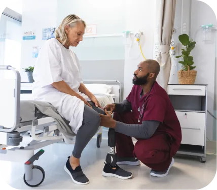
    Orthopedic clinician assisting older patient during recovery after a subchondral femur or tibial fracture treated with Subchondroplasty

    Ready to take the next step toward recovery?

    Schedule a consultation today with an orthopedic specialist to discuss Subchondral femur/tibial fracture treatment and learn how Subchondroplasty can help you get back to a pain-free, active life.

  • Chondral Defect/Injury Treatment: Understanding Your Symptoms and Options with Arthroscopic Chondroplasty

    Chondral Defect/Injury Treatment: Understanding Your Symptoms and Options with Arthroscopic Chondroplasty

    Cartilage damage in the knee or other joints is more common than most people realize, especially among active adults. When this smooth, protective surface wears down or becomes injured, it’s known as a chondral defect or chondral injury.

    While it might start as mild discomfort, untreated cartilage damage can gradually lead to stiffness, swelling, and eventually joint degeneration. The good news is that modern minimally invasive techniques — such as Arthroscopic Chondroplasty — can effectively treat this condition, relieve pain, and restore mobility.

    [tabla_contenidos]

    What Is a Chondral Defect or Injury?

    A chondral defect is an area where the cartilage that lines the joint surface has been damaged. Cartilage doesn’t have its own blood supply, which means it doesn’t heal easily once injured. These defects are often caused by:

    • A sports injury or fall
    • Repetitive stress on the joint
    • Previous ligament or meniscus injuries
    • Natural wear and tear over time

    The knee is the most common joint affected, but the same type of injury can occur in the ankle, hip, or shoulder. Recognizing the problem early is key to preventing further degeneration or arthritis.

    Common Symptoms: How Does It Feels

    Patients with a chondral defect often describe:

    • Pain deep inside the joint, especially with activity
    • Swelling or stiffness after exercise or long rest periods
    • Locking, catching, or grinding sensations when moving
    • Instability or the feeling that the joint “gives way”

    These symptoms can vary depending on how large and deep the defect is. Even small defects can cause discomfort if they’re located in high-pressure areas of the joint.

    How is Condral Defect/Injury Diagnosed?

    Your orthopedic specialist will start with a physical examination, evaluating joint movement, tenderness, and swelling.

    To confirm the diagnosis and determine the extent of cartilage damage, imaging studies are used:

    • X-rays rule out bone or alignment issues.
    • MRI scans provide detailed images of the cartilage surface.
    • In some cases, an arthroscopic evaluation allows direct visualization inside the joint for the most accurate assessment.

    This combination of exams helps your surgeon tailor the most effective treatment plan for your specific situation.

    Treatment Options

    Treatment for a chondral defect depends on the size, location, and severity of the injury — as well as your activity level and age.

    Non-Surgical Care

    Small or mild defects may be managed with:

    • Rest and physical therapy to strengthen muscles and reduce load on the joint
    • Anti-inflammatory medication for pain and swelling
    • Injection therapy, such as platelet-rich plasma (PRP) or hyaluronic acid

    These conservative treatments can provide relief but cannot restore the damaged cartilage. When symptoms persist, surgical intervention may be necessary.

    Surgical Treatment: Arthroscopic Chondroplasty

    When non-surgical options are no longer effective, Arthroscopic Chondroplasty offers a minimally invasive solution to repair and smooth damaged cartilage.

    What the Procedure Involves

    During this outpatient procedure, the surgeon inserts a small camera (arthroscope) and specialized instruments into the joint through tiny incisions. The damaged cartilage is trimmed, smoothed, and stabilized, creating a healthier surface that reduces friction and pain.

    The goal isn’t to regrow cartilage but to restore smoother joint motion and slow future wear. Because the surgery is done arthroscopically, it causes minimal disruption to surrounding tissues and allows for a faster recovery.

    When It’s Recommended

    Your orthopedic surgeon may recommend Arthroscopic Chondroplasty if you have:

    • Persistent pain or swelling that hasn’t improved with rest or therapy
    • Localized cartilage damage rather than widespread arthritis
    • Mechanical symptoms like catching, grinding, or locking
    • An active lifestyle and desire to return to sports or daily activities

    Benefits and Expected Outcomes

    • Minimally invasive – smaller incisions, less pain, and faster recovery
    • Effective pain relief – smoother surfaces reduce irritation
    • Improved mobility – restored joint motion and stability
    • High success rate – over 85–90% of patients experience lasting improvement
    • Prevents further damage by smoothing unstable cartilage edges

    Most patients notice significant relief within weeks and are able to resume daily activities much sooner than with open surgery.

    Long-Term Outlook

    When performed by an experienced orthopedic surgeon and followed by proper rehabilitation, Arthroscopic Chondroplasty can provide long-term pain relief and help delay or prevent the progression of arthritis. Regular follow-up visits ensure your recovery stays on track and your joint remains healthy.

    Why Choose a Specialist for Chondral Defect Treatment

    Cartilage injuries require precise diagnosis and expert treatment planning. An experienced orthopedic specialist who performs advanced arthroscopic procedures can identify the best approach for your specific joint and lifestyle goals.

    By combining minimally invasive surgery with personalized rehabilitation, the aim is to help you return to your normal activities — stronger, pain-free, and confident in your movement.

    Take the Next Step Toward Recovery

    Living with persistent joint pain isn’t something you have to accept. If you’ve been diagnosed with a chondral defect or cartilage injury, consult an orthopedic specialist experienced in Arthroscopic Chondroplasty.

    Orthopedic specialist examining a patient’s knee during evaluation for chondral defect and cartilage injury treatment

    With the right care, you can expect smoother movement, lasting relief, and a faster return to the activities you love.

  • Understanding End Stage Osteoarthritis: Symptoms, Diagnosis, and Treatment Options

    Understanding End Stage Osteoarthritis: Symptoms, Diagnosis, and Treatment Options

    Living with joint pain can be life-changing. For many adults, the pain and stiffness caused by osteoarthritis gradually worsen over time. When the joint cartilage wears down completely, it leads to what doctors call End Stage Osteoarthritis — the most advanced form of arthritis.

    If you’ve been told you have End Stage Osteoarthritis, understanding your condition and knowing your treatment options is the first step toward getting back to a pain-free, active life.

    [tabla_contenidos]

    What Is End Stage Osteoarthritis?

    End Stage Osteoarthritis occurs when the protective cartilage in your joint has completely worn away, leaving bone rubbing against bone. This stage is typically associated with significant pain, stiffness, and loss of mobility.

    It most often affects the knee, hip, or shoulder joints — the major weight-bearing joints that endure years of daily stress.

    By this point, conservative treatments like medications, injections, or physical therapy may no longer provide enough relief. Fortunately, advanced surgical techniques now offer highly effective solutions that can restore function and comfort — especially with the latest generation of personalized joint replacement implants.

    Common Symptoms of End Stage Osteoarthritis

    Patients with end stage osteoarthritis often describe a steady decline in their ability to move comfortably or perform daily activities. Some of the most common symptoms include:

    • Severe, constant joint pain, even at rest or during the night
    • Significant stiffness, especially after sitting or sleeping
    • Swelling and warmth around the joint
    • Grinding or clicking sensations during movement
    • Reduced range of motion and difficulty walking or bending
    • Visible joint deformity in more advanced cases

    These symptoms can be physically limiting and emotionally draining. However, modern orthopedic treatments can dramatically improve comfort and quality of life, even in advanced disease stages.

    How Is End Stage Osteoarthritis Diagnosed?

    Diagnosing end stage osteoarthritis involves a combination of clinical evaluation and imaging studies.

    Your orthopedic specialist will:

    1. Review your symptoms and medical history – including how long you’ve had pain and what treatments you’ve tried.
    2. Perform a physical examination – to assess swelling, joint alignment, stability, and range of motion.
    3. Order imaging tests, such as:
      • X-rays, which show loss of joint space, bone spurs, and deformity.
      • MRI scans, if needed, to evaluate soft tissue and cartilage damage.

    Once the diagnosis is confirmed, your orthopedic surgeon will discuss the best path forward — which may include surgical intervention if the joint damage is too extensive for non-surgical care to be effective.

    Treatment Options for End Stage Osteoarthritis

    Treatment is tailored to each patient’s age, lifestyle, and activity goals. While surgery is often recommended for end stage disease, doctors typically begin with non-surgical approaches to manage symptoms whenever possible.

    Non-Surgical Treatments

    Before considering surgery, your doctor may recommend:

    • Activity modification – avoiding high-impact exercises that worsen pain
    • Physical therapy – to maintain joint flexibility and strength
    • Anti-inflammatory medications – for temporary pain relief
    • Joint injections – such as corticosteroids or hyaluronic acid to reduce inflammation

    If these methods no longer provide adequate relief, it may be time to explore joint replacement surgery — the most effective solution for long-term pain relief and mobility restoration in end stage arthritis.

    Surgical Treatment: Cemented vs. Cementless TKA Utilizing PSI Implants

    When osteoarthritis reaches its final stage, one of the most successful treatments is Total Knee Arthroplasty (TKA), also known as knee replacement surgery.

    Today’s advancements allow surgeons to choose between cemented and cementless fixation techniques, often enhanced by Patient-Specific Instrumentation (PSI) implants — a technology that personalizes your surgery for optimal fit and alignment.

    What Are PSI Implants?

    Patient-Specific Instrumentation (PSI) involves creating surgical guides and implants customized to your unique knee anatomy. Using detailed MRI or CT scans, advanced software maps the exact contours of your knee.

    This data allows your orthopedic surgeon to:

    • Design precise cutting guides for bone preparation
    • Select or custom-create implants that perfectly match your joint shape
    • Ensure ideal alignment and balance for smoother joint function

    The result is a more natural-feeling knee, improved implant longevity, and faster recovery.

    Cemented Total Knee Arthroplasty (TKA)

    In a cemented TKA, the artificial knee components are fixed to the bone using a special medical-grade bone cement.

    This technique has been used for decades and offers:

    • Immediate implant stability, allowing earlier weight-bearing
    • Proven long-term success rates, with implants lasting 15–20 years or more
    • Excellent results in patients with lower bone density or older adults

    Cementless Total Knee Arthroplasty (TKA)

    In cementless TKA, the implant is designed with a porous surface that allows your natural bone to grow into it, securing it biologically instead of with cement.

    This approach is ideal for:

    • Younger, active patients with good bone quality
    • Those seeking a longer-lasting implant fixation
    • Patients looking for a more natural bone–implant bond

    Benefits of Cementless TKA

    • Strong biological fixation that integrates with your bone over time
    • Reduced risk of cement-related issues
    • Potentially longer implant lifespan for active individuals

    Why PSI Implants Make a Difference

    Combining PSI technology with either cemented or cementless TKA ensures the implant is positioned precisely to your anatomy.

    Key benefits include:

    • Shorter surgery time and less bone removal
    • More accurate alignment, improving long-term outcomes
    • Better soft tissue balance, reducing pain and stiffness
    • Enhanced comfort and stability, helping your new knee feel more natural

    Studies show that PSI-assisted knee replacements can improve satisfaction and reduce revision rates, especially when performed by an experienced orthopedic specialist.

    Taking the Next Step Toward Pain-Free Living

    End Stage Osteoarthritis doesn’t have to mean the end of an active lifestyle. With today’s advanced surgical techniques — particularly Cemented or Cementless TKA Utilizing PSI Implants — patients are achieving life-changing results.

    Whether you’re just beginning to explore your options or ready to discuss surgery, an experienced orthopedic specialist can help determine which approach is right for you.

    Ready to Move Without Pain Again?

    If joint pain is limiting your life, don’t wait. Schedule a consultation with an orthopedic surgeon experienced in End Stage Osteoarthritis treatment and Cemented vs. Cementless TKA Utilizing PSI Implants.

    Elderly woman speaking with an orthopedic specialist about end stage knee osteoarthritis treatment options

    A personalized evaluation and modern treatment plan can help you regain comfort, confidence, and the freedom to move again — pain-free.

  • Understanding Medial Compartment Chondromalacia: Causes, Symptoms, and Modern Treatment Options

    Understanding Medial Compartment Chondromalacia: Causes, Symptoms, and Modern Treatment Options

    If you’ve been diagnosed with medial compartment chondromalacia, you’re not alone — and the good news is, there are effective ways to manage and even restore knee function. Many adults under 65 experience this condition, especially those who are active or have had previous knee injuries.

    The first step toward meaningful recovery is understanding the “why” behind your knee pain. In this article, we’ll explain what medial compartment chondromalacia is, what symptoms to watch for, how it’s diagnosed, and the most effective treatments available today — including a promising minimally invasive solution called the MISHA Knee Implantation.

    [tabla_contenidos]

    What Is Medial Compartment Chondromalacia?

    “Chondromalacia” refers to the softening or deterioration of the cartilage that cushions the joint. In the medial compartment (the inner part of the knee), this means the smooth cartilage covering the ends of the femur and tibia starts to wear down.

    Over time, this breakdown can cause friction, inflammation, and discomfort when walking, climbing stairs, or even standing for long periods.

    Why Early Diagnosis Matters

    Catching medial compartment chondromalacia early allows your orthopedic specialist to slow down or even reverse progression before the cartilage loss leads to arthritis or joint deformity. The earlier we act, the better the long-term outlook for joint preservation.

    Common Symptoms

    Patients often describe their knee pain as:

    • A dull, aching pain on the inner side of the knee
    • Stiffness or swelling, especially after activity or long periods of sitting
    • Grinding or clicking sensations when bending the knee
    • Pain when going up or down stairs
    • Loss of confidence in the knee — feeling like it may “give out”

    In the early stages, symptoms may come and go. As the cartilage damage progresses, pain becomes more persistent and can start to interfere with everyday activities.

    How Is Medial Compartment Chondromalacia Diagnosed?

    An orthopedic evaluation begins with a detailed physical exam, during which your doctor checks for tenderness, range of motion, and joint stability.

    Imaging Tests

    To confirm the diagnosis and assess cartilage damage, imaging is typically performed:

    • X-rays: Show bone alignment and joint space narrowing.
    • MRI: Provides a clear view of cartilage, meniscus, and soft tissue health.
    • Diagnostic arthroscopy (in rare cases): A minimally invasive camera procedure to directly visualize the joint if needed.

    These tests help your surgeon determine both the severity of cartilage wear and whether it’s isolated to the medial compartment or involves other areas of the knee.

    Treatment Options for Medial Compartment Chondromalacia

    Treatment depends on how advanced the cartilage wear is and how much it’s affecting your daily life.

    Non-Surgical Treatments

    In the early stages, many patients benefit from:

    • Activity modification: Avoiding deep squats, high-impact exercise, and repetitive kneeling.
    • Physical therapy: Focused on strengthening the quadriceps and improving knee alignment.
    • Anti-inflammatory medications or injections: Reduce pain and swelling.
    • Weight management: Reduces stress on the inner knee joint.

    However, when pain persists despite these measures — and imaging shows cartilage loss in the medial compartment — your surgeon may discuss MISHA Knee Implantation as an advanced, joint-preserving option.

    MISHA Knee Implantation: A Modern Solution for Medial Compartment Chondromalacia

    What Is the MISHA Knee Implant?

    The MISHA Knee Implant (Medial Implantable Shock Absorber) is a new generation of minimally invasive joint preservation technology designed to relieve knee pain caused by medial compartment chondromalacia and early osteoarthritis.

    Instead of replacing the joint or cutting bone, the MISHA implant acts as a shock absorber placed on the inner side of the knee. It reduces the load on the damaged cartilage while allowing your knee to move naturally.

    Think of it as adding a cushion that takes pressure off the worn cartilage — allowing pain relief, improved mobility, and cartilage protection without resorting to joint replacement.

    How the Procedure Works

    The MISHA Knee Implantation procedure is performed through a small incision on the inside of the knee.

    • The surgeon carefully positions the implant between the femur and tibia bones.
    • It’s secured to the bone using a minimally invasive technique — no bone cutting or joint resurfacing is needed.
    • The implant immediately begins to absorb excess load from the inner knee, reducing pressure by up to 30%.

    The surgery typically takes less than one hour, and patients can often walk the same day.

    Who Is a Candidate for MISHA Knee Implantation?

    This treatment is best suited for adults who:

    • Have isolated medial compartment pain or cartilage damage
    • Still have healthy cartilage in other parts of the knee
    • Want to avoid or delay partial or total knee replacement
    • Lead an active lifestyle and wish to maintain mobility

    Your orthopedic specialist will evaluate your imaging and symptoms to confirm if MISHA is right for you.

    Benefits of MISHA Knee Implantation

    • Joint preservation: Keeps your natural bone and cartilage intact.
    • Minimally invasive: Smaller incision and faster recovery compared to traditional surgeries.
    • Immediate pain relief: Many patients report reduced pain within days.
    • Quick return to activity: Walking and light activity usually resume within weeks.
    • Reversible: The implant can be removed if needed, preserving future surgical options.

    Clinical studies have shown that the MISHA Knee Implant can significantly improve pain scores, function, and activity level, while delaying the need for knee replacement surgery in many patients.

    Long-Term Outlook and Success Rates

    The MISHA Knee Implant has shown high satisfaction rates among patients who wish to stay active without undergoing joint replacement. Studies demonstrate that it can reduce medial compartment pressure, relieve pain, and preserve cartilage health for years after surgery.

    For younger and middle-aged adults, it serves as a bridge therapy — providing real relief while keeping future surgical options open if the condition progresses.

    When to See an Orthopedic Specialist

    If you’ve been struggling with inner knee pain, stiffness, or swelling that isn’t improving with rest or therapy, it’s time to speak with an orthopedic specialist. Early evaluation allows you to explore less invasive, effective treatments like MISHA Knee Implantation before cartilage damage becomes severe.

    The Bottom Line

    Medial compartment chondromalacia can be frustrating, but it doesn’t have to limit your life. Thanks to modern treatments like the MISHA Knee Implant, patients now have a minimally invasive way to restore function, relieve pain, and stay active.

    Orthopedic specialist assessing an older adult’s knee during a physical therapy evaluation for medial compartment chondromalacia

    If you’re ready to explore your options, schedule a consultation with an experienced orthopedic surgeon today. The sooner you take action, the sooner you can return to the active, pain-free lifestyle you deserve.

  • Patellar Tendon/Quad Tendon Rupture Treatment: Understanding Your Injury and How Surgery Can Help You Recover Fully

    Patellar Tendon/Quad Tendon Rupture Treatment: Understanding Your Injury and How Surgery Can Help You Recover Fully

    A patellar tendon or quadriceps tendon rupture is a serious injury that affects the connection between your thigh muscles and your kneecap (patella), or between the kneecap and your shinbone (tibia). These tendons are essential for straightening the knee, walking, climbing stairs, and even standing up from a chair.

    When one of these tendons tears, the leg can lose its ability to extend properly — making simple movements extremely difficult or even impossible. While this injury can happen to anyone, it most often affects active adults under 65 who experience a sudden jump, fall, or trauma to the knee. Early diagnosis and treatment are crucial to restore full knee function and avoid long-term complications like weakness, stiffness, or instability.

    [tabla_contenidos]

    How to Recognize a Patellar or Quadriceps Tendon Rupture

    A tendon rupture in the front of the knee often presents very suddenly. Patients typically describe a sharp “pop” or tearing sensation, followed by immediate pain and difficulty moving the leg.

    Common symptoms include:

    • Sudden, severe pain just above (quadriceps tendon) or below (patellar tendon) the kneecap
    • Swelling and bruising around the front of the knee
    • A visible gap or indentation where the tendon has torn
    • Inability to straighten the knee or lift the leg when lying down
    • Difficulty walking or bearing weight on the injured leg
    • The kneecap may appear higher or lower than normal, depending on which tendon is affected

    If you experience these symptoms after a fall or sports injury, it’s important to seek medical attention right away. Early evaluation greatly improves the success of treatment and recovery.

    How Doctors Confirm the Injury

    Your orthopedic specialist will begin by asking about how the injury occurred and performing a physical examination. During the exam, your doctor will assess your ability to straighten the knee, check for swelling, and feel for any gaps in the tendon.

    To confirm the diagnosis and determine the severity of the tear, imaging tests are usually ordered:

    • X-rays can show changes in the position of the kneecap (too high or too low).
    • MRI (Magnetic Resonance Imaging) is the most accurate test for identifying a partial or complete tendon rupture and assessing damage to surrounding structures.
    • Ultrasound may also be used to visualize the tendon tear in real-time.

    Accurate diagnosis helps your surgeon decide whether non-surgical treatment is possible or if surgery is required to restore knee function.

    Treatment Options for Patellar/Quad Tendon Rupture

    Non-Surgical Treatment (For Partial Tears Only)

    Partial tears or minor strains may sometimes be treated without surgery, especially if the tendon is still partially intact. Treatment usually includes:

    • Immobilization with a knee brace or cast to keep the leg straight
    • Physical therapy after several weeks to regain motion and strength
    • Gradual return to normal activities over 2–3 months

    However, in most cases — particularly complete ruptures — non-surgical options cannot restore normal function. That’s when surgical repair becomes necessary.

    Surgical Treatment: Open Patellar Tendon/Quad Tendon Repair

    When the tendon is fully torn, the most effective solution is a surgical procedure called Open Patellar Tendon/Quad Tendon Repair. This operation directly repairs the torn tendon and restores the natural connection between your muscles, kneecap, and leg bone.

    When Surgery Is Recommended

    Surgery is strongly recommended when:

    • The tendon is completely ruptured (confirmed by MRI)
    • You are unable to straighten your leg
    • The kneecap is displaced from its normal position
    • You want to regain full knee function and return to sports or active work

    Delaying surgery can make the repair more difficult, as the tendon and muscles may shorten or scar down. Ideally, the repair should be performed within a few weeks of injury for the best results.

    How Open Patellar Tendon/Quad Tendon Repair Works

    During this procedure, your orthopedic surgeon makes a small incision over the front of the knee to carefully expose the torn tendon. The damaged ends are identified and reattached using strong surgical sutures or anchors that secure the tendon back to the kneecap or bone.

    Depending on the injury:

    • Patellar tendon repairs involve reattaching the tendon to the bottom of the kneecap.
    • Quadriceps tendon repairs involve reattaching the tendon to the top of the kneecap.

    In some cases, if the tissue is weak or retracted, the surgeon may use reinforcement with grafts or synthetic materials to strengthen the repair. The procedure typically takes less than 90 minutes and is done under regional or general anesthesia.

    Benefits of Open Patellar Tendon/Quad Tendon Repair

    Patients can expect excellent outcomes with timely surgery and proper rehabilitation. The main benefits include:

    • Restoration of knee strength and stability
    • Improved mobility and ability to walk normally again
    • Low risk of re-rupture when performed by an experienced orthopedic surgeon
    • Return to sports and active lifestyle within several months
    • High patient satisfaction — most people regain nearly full range of motion

    Clinical studies show that over 90% of patients recover good to excellent function after surgical repair when combined with structured rehabilitation.

    Success Rates and Long-Term Outlook

    The long-term outlook after Open Patellar Tendon/Quad Tendon Repair is excellent. When performed promptly and followed by a dedicated rehab program, the success rate exceeds 90%, and most patients return to their prior level of activity.

    Complications such as stiffness, weakness, or re-rupture are uncommon and can usually be minimized through early motion, consistent physical therapy, and follow-up with your orthopedic specialist.

    Take the First Step Toward Full Recovery

    A patellar or quadriceps tendon rupture is a serious injury, but with expert surgical care, full recovery is absolutely achievable. The key is early diagnosis and timely treatment by an experienced orthopedic specialist.

    If you’re struggling with knee pain, weakness, or difficulty straightening your leg after an injury, don’t wait — proper evaluation and treatment can make all the difference.

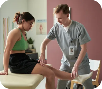
    Orthopedic specialist examining a patient’s knee during evaluation for a patellar or quadriceps tendon rupture

    Schedule a consultation with an orthopedic surgeon to discuss whether Open Patellar Tendon/Quad Tendon Repair is right for you. With the right surgical approach and personalized rehabilitation, you can confidently return to an active, pain-free life.